
Cystoscopy
Cystoscopy is a diagnostic and therapeutic procedure used to examine the bladder and urethra. It provides a direct visual assessment of the lower urinary tract, aiding in the diagnosis and treatment of various conditions.
Key Takeaways
- Cystoscopy involves inserting a thin tube with a camera into the urethra to visualize the bladder.
- It is used to diagnose and treat conditions like urinary tract infections, bladder stones, and tumors.
- The procedure can be performed with local or general anesthesia, depending on its scope.
- Risks are generally low but can include infection or temporary discomfort.
- Recovery is typically quick, with most individuals resuming normal activities within a day.
What is Cystoscopy?
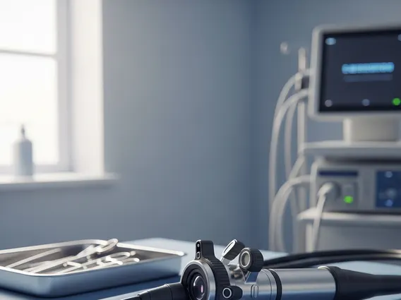
Cystoscopy is a medical procedure allowing a urologist to examine the lining of the bladder and the urethra. A thin, flexible or rigid tube called a cystoscope, equipped with a light and a camera, is inserted into the urethra and advanced into the bladder. This provides a direct visual inspection of the internal structures for abnormalities, crucial for diagnosing and monitoring various urological conditions.
This procedure, often referred to as a cystoscopy procedure, offers detailed images superior to external imaging techniques alone. It helps identify issues like inflammation, stones, tumors, or strictures within the urinary tract. The scope can also accommodate small instruments for minor surgical procedures, such as taking tissue samples (biopsies) or removing small stones.
How is a Cystoscopy Performed?
Performing a cystoscopy typically involves several steps for patient comfort and diagnostic accuracy. Patients may provide a urine sample beforehand to check for infection. The patient lies on their back with knees bent and feet in stirrups.
Here is a general overview of the procedure:
- Anesthesia: A local anesthetic gel is applied to the urethra for numbing. For more complex procedures or anxious patients, sedation or general anesthesia may be used.
- Insertion: The lubricated cystoscope is gently inserted into the urethra and advanced to the bladder.
- Visualization: Sterile saline solution distends the bladder for a clearer view of its wall. The urologist then examines the urethra and bladder lining.
- Intervention (if needed): Small instruments can be passed through the cystoscope to collect tissue samples, remove small growths, or treat certain conditions.
- Removal: After examination or intervention, saline is drained, and the cystoscope is carefully removed.
Cystoscopy Uses, Risks, and Recovery
Cystoscopy uses and risks are important considerations. It is primarily used for diagnosing and monitoring conditions affecting the bladder and urethra. Common applications include investigating blood in the urine (hematuria), recurrent urinary tract infections, painful urination, or overactive bladder symptoms. It is also crucial for detecting bladder stones, tumors, or blockages, and for monitoring patients with a history of bladder cancer.
While generally safe, potential risks are usually minor and temporary:
- Urinary tract infection (UTI)
- Temporary discomfort or burning during urination
- Small amount of blood in the urine
- Bladder spasm or urgency
Serious complications, such as bladder perforation or severe bleeding, are rare. Antibiotics may be administered to reduce infection risk.
Regarding cystoscopy recovery time, most individuals experience a quick recovery. Mild discomfort or burning during urination and slight blood in the urine are common for a day or two post-procedure. Drinking plenty of fluids can help alleviate irritation. Most patients can resume normal activities within 24 hours. If sedation was used, driving should be avoided for the rest of the day. Patients should contact their doctor for severe pain, heavy bleeding, fever, or inability to urinate.


