Computerized Tomography
Computerized Tomography (CT) is an advanced medical imaging technique that utilizes a series of X-ray images taken from different angles around the body. These images are then processed by a computer to create detailed cross-sectional slices of bones, blood vessels, and soft tissues inside the body.
What is Cancer
- CT scans combine X-rays and computer processing to produce highly detailed, cross-sectional images of the body.
- The technology is vital for diagnosing a wide range of conditions, from complex bone fractures to internal organ diseases and cancers.
- A CT scan works by rotating an X-ray source and detectors around the patient, collecting data that a computer reconstructs into images.
- Significant benefits of CT scans include their ability to provide superior detail of soft tissues and organs compared to conventional X-rays, aiding in rapid and accurate diagnosis.
What is Computerized Tomography?
What is Computerized Tomography? Often referred to simply as a CT scan, it is a non-invasive diagnostic imaging procedure that uses specialized X-ray equipment and sophisticated computers to generate multiple images or pictures of the inside of the body. Unlike traditional X-rays, which produce a single, flat image, CT scans create detailed, cross-sectional views, much like looking at slices of bread from a loaf. This allows medical professionals to visualize internal structures, including bones, soft tissues, and blood vessels, with exceptional clarity and depth.
How CT Scans Work
Understanding how does a CT scan work involves appreciating the interplay between X-ray technology and advanced computing. The process is designed to capture comprehensive internal views of the body quickly and effectively.
The Imaging Process
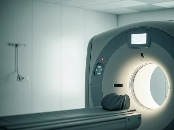
During a CT scan, the patient lies on a motorized table that slides into a large, doughnut-shaped machine called a gantry. Inside the gantry, an X-ray tube rotates rapidly around the patient, emitting a narrow beam of X-rays. On the opposite side of the patient, a series of detectors simultaneously measure the X-rays that pass through the body. As the X-ray tube and detectors rotate, they capture hundreds of thousands of individual X-ray measurements from various angles. A powerful computer then processes this vast amount of data, reconstructing it into detailed cross-sectional images, or “slices,” of the body. These slices can be viewed individually or combined to create three-dimensional (3D) representations of organs and structures.
Patient Preparation & Safety
Patient preparation for a CT scan is generally minimal but can vary depending on the area being examined. Patients may be asked to fast for a few hours before the scan, remove metal objects, or wear a hospital gown. In some cases, a contrast agent, administered orally or intravenously, may be used to enhance the visibility of specific tissues or blood vessels. Regarding safety, CT scans involve exposure to ionizing radiation. However, modern CT scanners are designed to minimize radiation dose while optimizing image quality. Medical professionals carefully weigh the diagnostic benefits against the potential risks, ensuring that the procedure is justified and performed safely. According to the American College of Radiology, the diagnostic benefits of CT scans typically outweigh the small potential risks of radiation exposure when medically indicated.
Uses and Benefits of CT Imaging
CT imaging has revolutionized medical diagnostics due to its versatility and the detailed information it provides. The uses of computerized tomography span a wide range of medical specialties, making it an indispensable tool in modern healthcare.
Common Diagnostic Applications
CT scans are frequently used to diagnose and monitor numerous conditions. They are highly effective in detecting bone fractures, especially complex ones involving the spine or pelvis, and in assessing internal injuries following trauma. For soft tissues, CT scans can identify tumors, infections, blood clots, and internal bleeding. They play a crucial role in diagnosing conditions affecting the brain, lungs, heart, abdominal organs, and blood vessels. Furthermore, CT imaging is often utilized to guide procedures such as biopsies, where a needle is precisely directed to collect tissue samples, and in planning radiation therapy for cancer treatment. This broad utility makes CT scans a cornerstone in emergency medicine, oncology, and many other diagnostic fields.
Advantages Over X-rays
The benefits of CT scans significantly extend beyond those offered by conventional X-rays. While X-rays provide a two-dimensional view, CT scans offer detailed cross-sectional images that reveal the precise size, shape, and location of abnormalities. This superior detail is particularly advantageous for visualizing soft tissues, such as organs, muscles, and blood vessels, which are often difficult to assess with standard X-rays. CT scans can also generate 3D reconstructions, providing a comprehensive anatomical overview that aids surgeons in planning complex procedures. Moreover, CT scans are generally faster than other advanced imaging techniques like MRI, making them invaluable in emergency situations where rapid diagnosis is critical for patient care. This combination of speed, detail, and versatility positions CT as a powerful diagnostic tool.