Computer Aided Detection System
A Computer Aided Detection System (CAD) represents a significant advancement in medical imaging, designed to assist healthcare professionals in identifying subtle abnormalities that might otherwise be overlooked. These systems play a crucial role in enhancing the accuracy and efficiency of diagnostic procedures across various medical specialties.
Key Takeaways
- A Computer Aided Detection System (CAD) is a technology that uses artificial intelligence to analyze medical images and highlight potential areas of concern for radiologists.
- CAD systems function by processing digital images, applying algorithms to detect patterns indicative of disease, and marking these regions for further review.
- They serve as a valuable second opinion, helping to improve the sensitivity of screenings and reduce the likelihood of missed diagnoses.
- Key benefits include enhanced diagnostic accuracy, earlier disease detection, and increased efficiency in image interpretation for medical professionals.
What is a Computer Aided Detection (CAD) System?
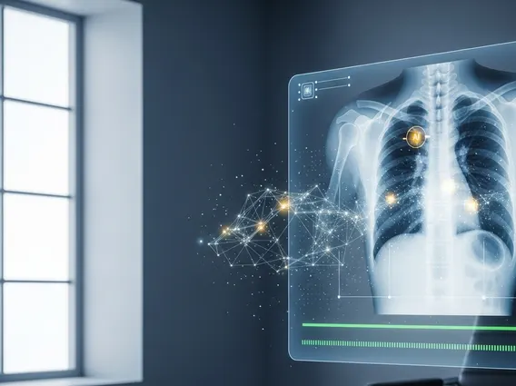
A Computer Aided Detection System (CAD) is an advanced technology used in medical imaging to assist radiologists and other medical professionals in interpreting diagnostic scans. These systems leverage sophisticated algorithms and artificial intelligence to analyze images, such as mammograms, CT scans, or X-rays, and identify potential areas of concern that may indicate the presence of disease. The primary goal of a CAD system is to act as a “second reader,” drawing attention to subtle findings that might be missed during a human review, thereby improving diagnostic accuracy.
The computer aided detection system explanation involves its ability to process vast amounts of image data, learning to recognize patterns associated with various conditions, such as tumors or lesions. For instance, in mammography, CAD systems are trained on large datasets of breast images to detect microcalcifications or masses that could be indicative of breast cancer. This technology does not make a diagnosis itself but rather serves as a valuable tool to augment the human eye, providing an objective analysis that complements the expertise of the interpreting physician.
How Computer Aided Detection Works
How computer aided detection works involves a multi-step process that integrates seamlessly into the medical imaging workflow. Initially, a digital medical image is acquired, such as a mammogram or a chest X-ray. This image is then fed into the CAD software. The system employs complex algorithms to analyze the image pixel by pixel, searching for specific features, textures, and patterns that have been previously identified as potential indicators of abnormalities.
The CAD software is trained using extensive databases of both normal and abnormal images. Through machine learning, it learns to differentiate between benign structures and suspicious findings. Once the analysis is complete, the system highlights or marks any areas it identifies as potentially abnormal directly on the image. These markers could be circles, arrows, or other visual cues. The radiologist then reviews the original image along with the CAD-generated marks. This allows the radiologist to pay closer attention to the flagged areas, making an informed decision about whether these findings are truly significant or merely artifacts. This collaborative approach combines the computational power of AI with the critical judgment and experience of a human expert.
Benefits of Computer Aided Detection Systems
The benefits of computer aided detection systems are substantial, contributing significantly to improved patient care and diagnostic efficiency. One of the most critical advantages is the enhancement of diagnostic accuracy. By providing a “second pair of eyes,” CAD systems can help radiologists detect subtle abnormalities that might be overlooked due to fatigue, distraction, or the sheer volume of images requiring interpretation. This is particularly vital in early disease detection, where even small lesions can be critical indicators.
According to the American Cancer Society, early detection of breast cancer through mammography, often aided by CAD, significantly improves treatment outcomes and survival rates. For example, the 5-year relative survival rate for localized breast cancer is 99% (American Cancer Society, 2023). CAD systems contribute to this by increasing the sensitivity of screening programs.
Other key benefits include:
- Increased Radiologist Efficiency: CAD systems can help streamline the reading process by quickly identifying areas of interest, allowing radiologists to focus their attention more effectively.
- Reduced False Negatives: By flagging suspicious areas, CAD can help reduce the rate of missed diagnoses, ensuring that fewer treatable conditions go undetected.
- Consistency in Interpretation: CAD offers a consistent, objective analysis, which can help standardize image interpretation across different radiologists and institutions.
- Support for Complex Cases: In cases with dense tissue or ambiguous findings, CAD can provide additional insights, aiding in more confident diagnostic decisions.
These systems ultimately empower healthcare providers to deliver more precise and timely diagnoses, leading to better patient outcomes.