
Comedo Carcinoma
Comedo Carcinoma is a specific type of ductal carcinoma in situ (DCIS) of the breast, characterized by distinct pathological features. Understanding this condition is crucial for accurate diagnosis and effective management in oncology.
Key Takeaways
- Comedo carcinoma is a high-grade form of ductal carcinoma in situ (DCIS) of the breast.
- It is characterized by central necrosis within the affected ducts, often visible on mammography as microcalcifications.
- Symptoms are typically subtle or absent, with diagnosis often occurring through screening mammograms.
- Diagnosis relies on biopsy and histopathological examination to confirm the presence of necrotic cells.
- Treatment primarily involves surgical excision, often followed by radiation therapy, to prevent progression to invasive cancer.
What is Comedo Carcinoma?
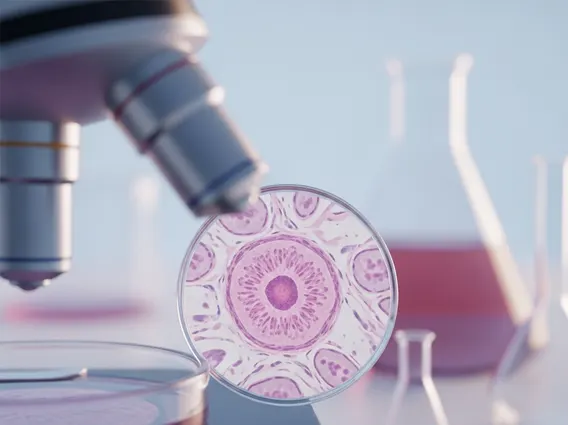
Comedo Carcinoma is a specific, high-grade subtype of ductal carcinoma in situ (DCIS) of the breast. DCIS represents non-invasive breast cancer where abnormal cells are confined to the milk ducts and have not spread to surrounding breast tissue. What distinguishes comedo carcinoma from other forms of DCIS is the presence of central necrosis (dead cells) within the affected ducts, often accompanied by calcifications. These necrotic cells can sometimes be “expressed” from the duct, resembling comedones (blackheads), hence the name. DCIS, in general, accounts for approximately 20-25% of all newly diagnosed breast cancers in the United States, according to the American Cancer Society, highlighting its prevalence as a non-invasive precursor.
Symptoms, Causes, and Diagnosis
Comedo carcinoma symptoms and causes are often subtle, making early detection challenging without routine screening. Many individuals with this condition experience no noticeable symptoms, as the abnormal cells are confined within the breast ducts. When symptoms do occur, they might include a palpable lump or thickening in the breast, or occasionally, nipple discharge. However, the most common way Comedo Carcinoma is detected is through screening mammography, which often reveals characteristic microcalcifications. These calcifications are a result of the necrotic debris within the ducts.
The exact causes of comedo carcinoma, like other forms of breast cancer, are multifactorial and not fully understood. They are believed to involve a combination of genetic predispositions, hormonal influences, and environmental factors that lead to uncontrolled cell growth within the breast ducts. Risk factors for developing DCIS, including the comedo subtype, are similar to those for invasive breast cancer, such as increasing age, a personal or family history of breast cancer, certain genetic mutations (e.g., BRCA1/2), and dense breast tissue.
Diagnosing comedo carcinoma typically begins with imaging studies. Mammography is crucial, as it can identify the suspicious microcalcifications often associated with this subtype of DCIS. If suspicious findings are noted, further imaging like ultrasound or MRI may be performed. The definitive diagnosis, however, requires a biopsy, usually a core needle biopsy, to obtain tissue samples. These samples are then examined under a microscope by a pathologist, who can confirm the presence of high-grade atypical cells with central necrosis, characteristic of comedo carcinoma. This histopathological examination is essential for distinguishing it from other types of DCIS or benign conditions.
The diagnostic process for comedo carcinoma typically involves several key steps:
- Screening Mammography: Often the first line of detection, revealing suspicious microcalcifications.
- Diagnostic Imaging: Further evaluation with targeted mammography, ultrasound, or MRI to characterize abnormalities.
- Biopsy: A core needle biopsy is performed to extract tissue samples from the suspicious area.
- Histopathological Examination: A pathologist analyzes the tissue to confirm the presence of high-grade ductal cells with central necrosis.
Comedo Carcinoma Treatment Options
Comedo carcinoma treatment options primarily focus on removing the abnormal cells and preventing the progression to invasive breast cancer. Since it is a non-invasive condition, the goal is local control. The standard approach typically involves surgery, often followed by radiation therapy.
Surgical intervention is the cornerstone of treatment. This usually involves a lumpectomy (breast-conserving surgery), where the tumor and a margin of healthy tissue are removed. The aim is to achieve clear margins, meaning no cancer cells are found at the edges of the removed tissue. In some cases, if the area of DCIS is extensive or if clear margins cannot be achieved with lumpectomy, a mastectomy (removal of the entire breast) may be recommended. Sentinel lymph node biopsy is generally not performed for pure DCIS, including comedo carcinoma, unless there are concerns about microinvasion or if a mastectomy is planned.
Following surgery, radiation therapy is often recommended, especially after a lumpectomy, to reduce the risk of recurrence in the treated breast. This adjuvant therapy targets any remaining microscopic cancer cells that might not have been removed during surgery. For certain patients, endocrine therapy (e.g., tamoxifen) may be considered if the DCIS cells are hormone receptor-positive, further reducing the risk of future breast cancer events. The specific treatment plan is individualized based on factors such as the size and grade of the DCIS, hormone receptor status, and patient preferences, always aiming to minimize recurrence while preserving quality of life.


