Chest X Ray
A Chest X Ray is a common imaging test that uses a small amount of radiation to create pictures of the chest, including the heart, lungs, blood vessels, airways, and the bones of the chest and spine. It’s a quick and non-invasive way for doctors to assess the health of these vital organs and structures.
Key Takeaways
- A Chest X Ray uses X-rays to produce images of the internal chest structures, aiding in the diagnosis of various conditions.
- It’s a quick, non-invasive procedure vital for assessing lung, heart, and bone health.
- The process involves brief exposure to radiation, with images captured on a detector.
- Doctors use chest X-rays to diagnose conditions like pneumonia, heart failure, and fractures.
- Interpreting chest x-ray results requires medical expertise to identify abnormalities and guide treatment.
What is a Chest X-Ray?
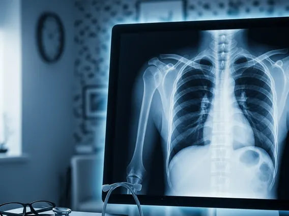
A Chest X Ray, also known as a chest radiograph, is a fundamental diagnostic tool in medicine. It is an imaging technique that utilizes X-rays to generate two-dimensional images of the organs and structures inside the chest cavity. This includes the lungs, heart, large blood vessels, diaphragm, and the bones of the chest wall and spine. The primary purpose of understanding what is Chest X Ray is to help physicians detect, diagnose, and monitor various medical conditions affecting these areas, providing crucial insights into a patient’s health without invasive procedures.
How Does a Chest X-Ray Work?
Understanding how does a chest x-ray work involves grasping the principles of radiation. During the procedure, a machine sends a controlled beam of X-rays through the patient’s chest. Different tissues absorb X-rays at varying rates. For instance, dense structures like bones absorb more X-rays and appear white on the image. Soft tissues such as the lungs, which are filled with air, absorb fewer X-rays and appear darker. The X-rays that pass through the body are captured by a special detector, either a photographic film or a digital sensor, which then creates the visual image. This rapid process typically takes only a few seconds.
Purpose and Procedure of a Chest X-Ray
A Chest X Ray serves a broad range of diagnostic purposes, making it one of the most frequently performed imaging tests globally. What is a chest x-ray used for includes diagnosing conditions such as pneumonia, tuberculosis, lung cancer, heart failure, emphysema, and fractured ribs. It can also help monitor the progression of diseases or the effectiveness of treatments.
The chest x-ray procedure information is straightforward and generally takes only a few minutes. Patients will typically be asked to remove any jewelry or metal objects that could interfere with the X-ray image. They will then stand or sit between the X-ray machine and a plate that contains the X-ray film or digital sensor. The radiographer will position the patient, usually requiring them to take a deep breath and hold it for a few seconds to ensure the lungs are fully expanded and the heart is clearly visible. Multiple views, such as a posterior-anterior (PA) view and a lateral view, are often taken to provide a comprehensive assessment of the chest structures. The exposure to radiation is minimal and considered safe for most individuals.
Interpreting Chest X-Ray Results
The process of interpreting chest x-ray results is performed by a radiologist, a medical doctor specializing in interpreting medical images. The radiologist examines the images for any abnormalities in the size, shape, and position of the heart, lungs, and other structures. They look for signs of infection, inflammation, fluid accumulation, tumors, fractures, or other indicators of disease. For example, pneumonia might appear as white patches (infiltrates) in the lungs, while an enlarged heart could suggest heart failure.
Once the radiologist has analyzed the images, they will provide a detailed report to the referring physician. The physician then discusses these findings with the patient, explaining what the results mean in the context of their symptoms and medical history. Further tests may be recommended based on the findings to confirm a diagnosis or guide treatment. It’s important to remember that a chest X-ray is one piece of the diagnostic puzzle, and its results are always considered alongside other clinical information.