
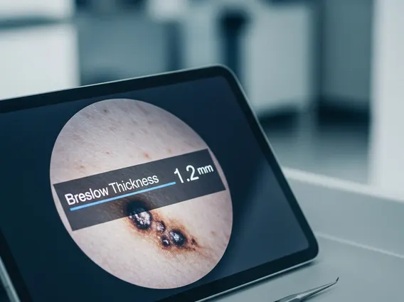
Breslow Thickness
Breslow thickness is a critical measurement used in the diagnosis and staging of melanoma, a serious form of skin cancer. It quantifies the vertical depth of the tumor, providing essential information for prognosis and guiding treatment decisions.
Key Takeaways
- Breslow thickness measures the vertical depth of melanoma from the top of the epidermis to the deepest tumor cell.
- It is the most important prognostic factor for localized melanoma, directly correlating with the risk of metastasis.
- The measurement is performed by a pathologist and is crucial for determining the melanoma’s stage.
- Different thickness levels guide subsequent treatment, including surgical margins and the need for sentinel lymph node biopsy.
What is Breslow Thickness?
What is Breslow Thickness refers to the measurement of the vertical depth of a melanoma tumor. This measurement is taken from the top of the granular layer of the epidermis (or the base of a superficial ulcer) down to the deepest identifiable invasive melanoma cell. Developed by Dr. Alexander Breslow in 1970, this metric has become the single most important prognostic factor for patients with localized melanoma, indicating the tumor’s potential to spread.
The concept behind Breslow thickness is straightforward: thinner melanomas are generally associated with a better prognosis, while thicker melanomas indicate a higher risk of metastasis and recurrence. This depth measurement helps clinicians understand the aggressiveness of the cancer and plan appropriate management strategies.
How is Breslow Thickness Measured?
The process of Breslow thickness measurement explained involves a pathologist examining a biopsy sample of the melanoma under a microscope. Using a calibrated ocular micrometer, the pathologist precisely measures the distance in millimeters (mm) from the epidermal surface (or the base of an ulceration, if present) to the deepest point of tumor invasion. This meticulous measurement is then recorded in the pathology report, forming a cornerstone of the patient’s diagnostic information.
Accuracy in this measurement is paramount, as even small differences can impact staging and treatment recommendations. The pathologist ensures that the measurement captures the true vertical extent of the tumor, providing a reliable basis for clinical decisions.
Significance in Melanoma Staging
The Breslow thickness significance in diagnosis cannot be overstated, as it is a primary determinant in the staging of melanoma according to the American Joint Committee on Cancer (AJCC) staging system. This system categorizes melanoma into stages (0 to IV), with Breslow thickness playing a pivotal role in Stage I and II classifications. For instance, a melanoma with a Breslow thickness of less than 1.0 mm without ulceration is typically considered Stage IA, whereas a thicker melanoma, especially with ulceration, would be classified into a higher stage.
Understanding Breslow thickness melanoma stages is crucial for both clinicians and patients. Thinner melanomas (e.g., less than 1.0 mm) are associated with a significantly lower risk of regional lymph node metastasis and distant spread. Conversely, melanomas with a thickness greater than 1.0 mm carry an increased risk, prompting further diagnostic procedures like sentinel lymph node biopsy.
Impact on Prognosis and Treatment
The impact of Breslow thickness on prognosis and treatment is profound. It directly correlates with survival rates and the likelihood of recurrence. For example, the 5-year survival rate for patients with thin melanomas (less than 1.0 mm) is generally very high, often exceeding 95%. However, for melanomas thicker than 4.0 mm, the 5-year survival rate can drop significantly, sometimes below 50%, especially if there is ulceration or lymph node involvement. (Source: National Cancer Institute SEER Program data, consistently shows this trend).
Treatment decisions are heavily influenced by this measurement. For thin melanomas, wide local excision with relatively small surgical margins is often sufficient. For thicker melanomas (typically >1.0 mm or with ulceration), wider surgical margins are recommended, and a sentinel lymph node biopsy may be performed to check for microscopic spread to nearby lymph nodes. The results of these evaluations, guided by Breslow thickness, determine the need for adjuvant therapies, such as immunotherapy or targeted therapy, to reduce the risk of recurrence and improve long-term outcomes.


