
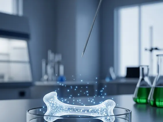
Bone Seeking Radioisotope
Bone seeking radioisotopes are specialized radioactive substances designed to target and accumulate within bone tissue. They play a crucial role in modern medicine, offering unique capabilities for both diagnosis and treatment of various bone-related conditions.
Key Takeaways
- Bone seeking radioisotopes are radioactive compounds that selectively accumulate in bone.
- Their mechanism relies on mimicking calcium or binding to bone mineral, concentrating radiation at specific sites.
- They are used for both diagnostic imaging (e.g., bone scans) and therapeutic interventions (e.g., pain palliation, cancer treatment).
- Common types include Technetium-99m for diagnostics and Strontium-89 or Radium-223 for therapy.
- These agents provide targeted delivery of radiation, minimizing impact on healthy tissues.
What is a Bone Seeking Radioisotope?
A bone seeking radioisotope is a radioactive pharmaceutical agent specifically engineered to localize within the skeletal system. These isotopes are designed to be absorbed by bone tissue, either by mimicking naturally occurring minerals like calcium or by binding to the bone matrix. This selective accumulation allows for the targeted delivery of radiation directly to areas of active bone metabolism, which can be indicative of disease or injury. Understanding what are bone seeking radioisotopes is fundamental to appreciating their utility in nuclear medicine, where their unique properties are harnessed for precise medical applications.
Mechanism of Bone Seeking Radioisotopes
The mechanism of how do bone seeking radioisotopes work primarily involves their affinity for bone mineral. Many of these agents are analogs of calcium or phosphate, which are integral components of hydroxyapatite, the primary mineral in bone. When introduced into the body, these radioisotopes circulate in the bloodstream and are preferentially incorporated into areas of active bone remodeling or increased osteoblastic activity. For instance, Technetium-99m-MDP (methylene diphosphonate) binds to the hydroxyapatite crystals on the surface of bone, particularly in regions where new bone is being formed or repaired. This selective uptake ensures that the emitted radiation is concentrated at the target site, allowing for imaging or therapeutic effect with minimal exposure to surrounding healthy tissues. The specific type of radiation emitted (gamma for imaging, beta or alpha for therapy) determines its application.
Types and Medical Applications
There are several types of bone seeking radioisotopes, each tailored for specific diagnostic or therapeutic purposes. The choice of radioisotope depends on the clinical objective, whether it’s to visualize bone abnormalities or to deliver targeted radiation for treatment. The uses of bone seeking radioisotopes are diverse and critical in oncology, orthopedics, and rheumatology, providing invaluable tools for managing a range of conditions.
| Radioisotope | Type of Radiation | Primary Use | Medical Application |
|---|---|---|---|
| Technetium-99m (Tc-99m) MDP | Gamma | Diagnostic | Bone scans for detecting fractures, infections, tumors, and metabolic bone diseases. It is the most widely used diagnostic agent, offering high resolution images of bone metabolism. |
| Strontium-89 (Sr-89) Chloride | Beta | Therapeutic | Pain palliation for metastatic bone cancer, particularly prostate and breast cancer. It mimics calcium and is selectively incorporated into areas of increased bone turnover, delivering localized radiation. |
| Samarium-153 (Sm-153) EDTMP | Beta and Gamma | Therapeutic & Diagnostic | Pain relief from bone metastases; the gamma emission also allows for post-treatment imaging, though its primary role is therapeutic. |
| Radium-223 (Ra-223) Dichloride | Alpha | Therapeutic | Treatment of metastatic castration-resistant prostate cancer with symptomatic bone metastases. Its potent alpha particles have a very short range, minimizing damage to surrounding healthy tissues while effectively targeting cancer cells in bone. |
These radioisotopes enable clinicians to non-invasively assess bone health, identify disease progression, and provide targeted treatment, significantly improving patient outcomes. The widespread adoption of these techniques underscores their importance; for instance, diagnostic bone scans are a cornerstone of nuclear medicine imaging, frequently performed globally to evaluate skeletal integrity and pathology, as highlighted by various professional nuclear medicine societies. This consistent use demonstrates their proven efficacy and safety in clinical practice.


