
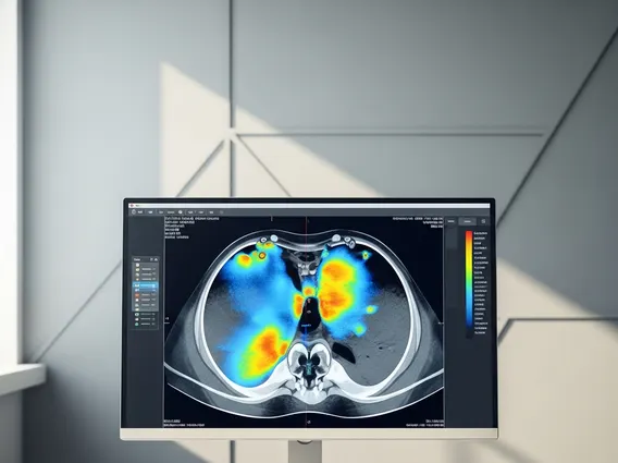
Bone Scan
A bone scan is a specialized diagnostic imaging test that provides detailed insights into the health and function of your bones. It is widely used to detect and evaluate various bone conditions that might not be visible through conventional imaging methods.
Key Takeaways
- A bone scan utilizes a small, safe amount of radioactive tracer to highlight areas of increased or decreased bone metabolism.
- Its primary purpose of a bone scan is to identify bone diseases, hidden fractures, infections, and the spread of certain cancers to the bones.
- The bone scan procedure explained involves an initial tracer injection, a waiting period for tracer uptake, and subsequent imaging with a specialized gamma camera.
- This diagnostic tool helps physicians make early diagnoses, monitor disease progression, and assess the effectiveness of treatments.
What is a Bone Scan and Its Purpose?
A bone scan is a nuclear medicine imaging test that helps physicians diagnose and track several types of bone diseases. It involves injecting a small amount of radioactive material, known as a tracer, into a vein. This tracer travels through the bloodstream and accumulates in areas of the bones that are undergoing rapid metabolism or repair. The areas where the tracer accumulates more intensely indicate increased bone activity, which can be a sign of various conditions such as inflammation, infection, or tumor growth. This technique is particularly sensitive to changes in bone metabolism, often detecting abnormalities earlier than standard X-rays.
The primary purpose of a bone scan is to identify bone abnormalities that might not be apparent on conventional X-rays. For instance, it can detect stress fractures, bone infections (osteomyelitis), and certain types of cancer that have spread to the bones (metastasis). According to the American Cancer Society, bone scans are frequently used in cancer staging to check for bone involvement, particularly for cancers like prostate, breast, and lung cancer, as bone metastases are common in these cases. This diagnostic tool is invaluable for early detection, assessing the extent of disease, and monitoring the progression of bone-related conditions, helping guide appropriate treatment strategies.
How a Bone Scan Works: The Procedure Explained
Understanding how bone scan works involves recognizing the role of the radioactive tracer and the sophisticated imaging equipment. Once injected, the tracer, typically a technetium-99m labeled phosphate compound, is absorbed by bone cells. Areas with higher metabolic activity, such as those with inflammation, infection, or rapid bone growth (like in healing fractures or tumors), will absorb more tracer. A special camera, known as a gamma camera, then detects the radiation emitted by the tracer in the bones and creates detailed images. These images, often called scintigrams, show “hot spots” (areas of increased tracer uptake) or “cold spots” (areas of decreased uptake), which correspond to abnormal bone activity. The intensity and pattern of tracer uptake provide crucial information for diagnosis.
The bone scan procedure explained typically involves a few key steps to ensure accurate results:
- Tracer Injection: A small, safe amount of radioactive tracer is injected into a vein, usually in the arm. This tracer is designed to target bone tissue and is quickly eliminated from the body over time.
- Waiting Period: There is typically a waiting period of 2-4 hours after the injection. During this time, the tracer travels throughout the body and accumulates in the bones. Patients are usually encouraged to drink plenty of water to help clear any unbound tracer from soft tissues, improving image clarity.
- Imaging: The patient lies comfortably on a table while a gamma camera moves slowly over their body, capturing images. This imaging process usually takes 30-60 minutes. In some cases, a three-phase bone scan might be performed, involving immediate images after injection, followed by images a few minutes later, and then the standard delayed images, to assess blood flow and soft tissue uptake more thoroughly.
- Interpretation: A nuclear medicine physician, specialized in interpreting these images, analyzes the patterns of tracer uptake. The results are then compiled into a report and shared with the referring physician to inform diagnosis and treatment planning.
This procedure is generally considered safe, with minimal radiation exposure comparable to a standard X-ray, and typically involves no significant side effects.


