
Birth Canal
The birth canal is a crucial anatomical pathway through which a baby passes during vaginal childbirth. Understanding its structure and function is essential for comprehending the physiological process of labor and delivery.
Key Takeaways
- The birth canal is the pathway for vaginal delivery, comprising the pelvis, cervix, vagina, and pelvic floor.
- Its primary function is to guide the baby from the uterus to the outside world during childbirth.
- During labor, the cervix dilates and the baby descends through the pelvic bones and soft tissues.
- The tissues of the birth canal demonstrate remarkable flexibility and stretching capabilities to accommodate the baby’s passage.
- The coordinated efforts of uterine contractions and maternal pushing facilitate the baby’s journey through this complex pathway.
What is the Birth Canal?
The term birth canal refers to the combined anatomical structures that form the passageway for a baby during vaginal delivery. This intricate pathway is designed to facilitate the journey from the mother’s uterus to the external environment. Understanding what is the birth canal involves examining its bony and soft tissue components, which work in concert to achieve childbirth.
Anatomy of the Birth Canal
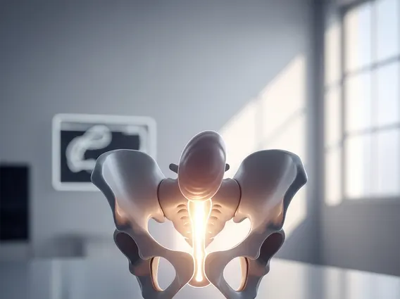
The birth canal anatomy and function are intrinsically linked, with each component playing a vital role. It primarily consists of the bony pelvis, the cervix, the vagina, and the pelvic floor muscles. The bony pelvis forms a rigid ring, defining the initial dimensions through which the baby must pass. The cervix, the lower part of the uterus, must dilate significantly to allow the baby’s head to enter the vagina. The vagina itself is a muscular, elastic tube that connects the uterus to the outside, and the pelvic floor muscles provide support and also need to relax and stretch during delivery.
Primary Function in Childbirth
The primary function of the birth canal is to provide a safe and efficient route for the baby’s passage during childbirth. This pathway guides the baby through a series of rotations and descents, adapting to the contours of the maternal pelvis. The coordinated actions of uterine contractions, which push the baby downwards, and the gradual yielding of the soft tissues of the birth canal, ensure the baby’s progression. This complex interplay is fundamental to the successful completion of a vaginal delivery.
How the Birth Canal Works During Labor
During labor, the birth canal undergoes a series of dynamic changes to accommodate the baby’s journey. The process involves precise coordination between maternal physiological responses and the baby’s movements, illustrating how birth canal works during labor to achieve delivery.
Stages of Dilation and Descent
The process begins with cervical effacement and dilation, where the cervix thins and opens. This is often measured in centimeters, with full dilation typically being 10 centimeters. As labor progresses, the baby’s head descends through the various planes of the bony pelvis. This descent involves molding of the baby’s head and specific rotations to navigate the narrowest parts of the pelvis. According to the World Health Organization (WHO), the active phase of labor, characterized by rapid cervical dilation, typically progresses at a rate of at least 1 cm per hour for first-time mothers. The baby’s descent is monitored by healthcare providers to ensure a safe progression through the birth canal.
Birth Canal Stretching and Flexibility
A remarkable aspect of how birth canal works during labor is its capacity for stretching and flexibility. The soft tissues of the vagina and pelvic floor are highly elastic, allowing them to expand significantly to accommodate the baby’s head and body. This process, known as birth canal stretching during delivery, is crucial for preventing injury to both mother and baby. Hormonal changes during pregnancy, particularly increased levels of relaxin, contribute to the softening and increased elasticity of ligaments and tissues, preparing the body for childbirth. The ability of these tissues to stretch and then return to their approximate pre-pregnancy state highlights the incredible adaptability of the female anatomy.


