Atypical Mole
An atypical mole, also known as a dysplastic nevus, is a common type of mole that can appear unusual and sometimes resemble melanoma. While generally benign, understanding their characteristics and the importance of monitoring them is crucial for skin health.
Key Takeaways
- Atypical moles (dysplastic nevi) are unusual moles with irregular features that can be mistaken for melanoma.
- They are not cancerous but indicate an increased risk of developing melanoma elsewhere on the body.
- Recognizing changes in size, shape, color, or border is essential for early detection.
- Regular self-skin exams and professional dermatological checks are vital for individuals with atypical moles.
- A biopsy is often required to definitively distinguish an atypical mole from melanoma.
What is an Atypical Mole (Dysplastic Nevus)?
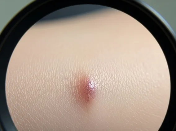
An atypical mole, medically termed a dysplastic nevus, is a benign (non-cancerous) skin growth that looks different from a common mole. These moles often have irregular features, making them challenging to distinguish from melanoma without professional evaluation. They tend to be larger than typical moles, often exceeding 6 millimeters in diameter, and can have indistinct or fuzzy borders. The color within an atypical mole may vary, presenting shades of tan, brown, and sometimes even pink or red. While most atypical moles remain benign, their presence is a significant indicator of an increased risk for developing melanoma, either within the atypical mole itself or on other areas of the skin. Therefore, comprehensive dysplastic nevus information emphasizes the need for vigilance and regular skin examinations.
Recognizing Atypical Mole Symptoms
Identifying atypical mole symptoms involves paying close attention to specific visual characteristics that deviate from those of common moles. While common moles are usually symmetrical, uniformly colored, and have distinct borders, atypical moles often exhibit irregularities. Dermatologists use the “ABCDE” rule as a guide for identifying suspicious lesions, which can also apply to monitoring atypical moles for changes:
- Asymmetry: One half of the mole does not match the other half.
- Border irregularity: The edges are ragged, notched, blurred, or poorly defined.
- Color variation: The mole has different shades of tan, brown, black, or sometimes areas of red, white, or blue.
- Diameter: The mole is typically larger than 6 millimeters (about the size of a pencil eraser), though melanoma can sometimes be smaller.
- Evolving: Any change in size, shape, color, elevation, or any new symptoms such as bleeding, itching, or tenderness.
It is crucial to regularly perform self-skin exams and consult a dermatologist if any of these symptoms are observed in an existing mole or if a new, unusual mole appears.
Atypical Mole vs. Melanoma: Key Differences
Distinguishing an atypical mole vs. melanoma is a critical aspect of skin cancer prevention, as they can share similar visual characteristics. The primary difference is that an atypical mole is a benign growth, meaning it is not cancerous, whereas melanoma is a malignant form of skin cancer that can spread if not treated early. While an atypical mole itself is not melanoma, it represents a precursor lesion and a marker for increased melanoma risk. According to the American Academy of Dermatology, individuals with five or more atypical moles have a 6- to 10-fold increased risk of developing melanoma compared with the general population.
Because of their similar appearances, a definitive diagnosis often requires a biopsy, where a small tissue sample is removed and examined under a microscope. Dermatologists use specialized tools like a dermatoscope to examine moles more closely, but only a biopsy can confirm whether a lesion is an atypical mole or melanoma. Regular professional skin examinations are essential for individuals with atypical moles to monitor for any changes that might indicate the development of melanoma.