Hamartoma
A hamartoma is a benign (non-cancerous) growth that occurs when cells and tissues develop in a disorganized manner in their normal location. These growths are typically discovered incidentally and often do not require intervention.
Key Takeaways
- Hamartoma is a benign, non-cancerous growth of normal tissue in an abnormal arrangement.
- Symptoms vary greatly depending on the hamartoma’s location and size, with many being asymptomatic.
- Causes are often unknown, though some are associated with genetic syndromes.
- Diagnosis typically involves imaging studies and sometimes a biopsy.
- Treatment ranges from observation to surgical removal, depending on symptoms and potential complications.
What is Hamartoma?
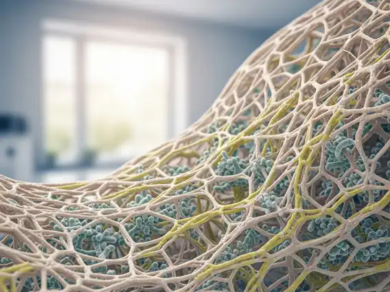
A Hamartoma refers to a focal malformation that resembles a tumor but is actually an overgrowth of mature cells and tissues that are normally found in the affected area. Unlike malignant tumors, hamartomas do not metastasize (spread) to other parts of the body. They are essentially developmental anomalies, where the constituent cells grow in a disorganized fashion, often forming a mass. While they can occur in almost any organ, common sites include the lungs, skin, brain, and kidneys. The size of a hamartoma can vary significantly, from microscopic to several centimeters, and their growth rate is generally slow or they may remain stable over time.
Understanding what is a hamartoma is crucial for distinguishing it from cancerous growths. These lesions are composed of the same types of cells as the surrounding tissue but are arranged abnormally. For instance, a hamartoma in the lung might contain a mix of cartilage, fat, and connective tissue, which are all normal components of the lung, but are present in an irregular, tumor-like formation. Their benign nature means they are generally not life-threatening, though their location and size can sometimes lead to functional impairment or symptoms.
Hamartoma Symptoms, Causes, and Types
The presentation of hamartoma symptoms and causes is highly variable, largely depending on the location and size of the growth. Many hamartomas are asymptomatic and are discovered incidentally during imaging for other conditions. When symptoms do occur, they are typically due to the mass effect of the hamartoma compressing surrounding tissues or interfering with organ function. For example, a hamartoma in the lung might cause coughing or shortness of breath, while one in the brain could lead to seizures or neurological deficits.
The exact causes of hamartomas are often unknown, but some are associated with specific genetic syndromes. For instance, tuberous sclerosis complex is linked to hamartomas in the brain, kidneys, and skin. Other hamartomas may arise sporadically without any clear genetic predisposition. Research continues to explore the molecular pathways involved in their development.
When considering types of hamartoma explained, they are generally classified by their location and the predominant tissue type involved. Some common types include:
- Pulmonary Hamartoma: The most common benign tumor of the lung, often composed of cartilage, connective tissue, and fat.
- Hypothalamic Hamartoma: A rare brain lesion that can cause gelastic seizures (laughing seizures) and precocious puberty.
- Renal Hamartoma (Angiomyolipoma): Often associated with tuberous sclerosis, these kidney growths contain fat, smooth muscle, and blood vessels.
- Skin Hamartoma: Can manifest as various skin lesions, such as epidermal nevi or connective tissue nevi.
- Breast Hamartoma: A benign lesion of the breast composed of glandular, fibrous, and adipose tissue, often presenting as a well-circumscribed mass.
Diagnosis and Treatment of Hamartoma
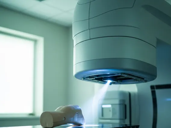
The diagnosis and treatment of hamartoma typically begins with imaging studies. Depending on the suspected location, this might include X-rays, computed tomography (CT) scans, magnetic resonance imaging (MRI), or ultrasound. These imaging techniques help to identify the presence, size, and characteristics of the mass. For instance, a pulmonary hamartoma often has a characteristic “popcorn” calcification pattern on CT scans. In many cases, imaging alone can strongly suggest a hamartoma, especially if it has typical features and remains stable over time.
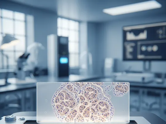
However, to definitively confirm the diagnosis and rule out malignancy, a biopsy may be necessary. This involves taking a small tissue sample from the growth for microscopic examination by a pathologist. Once a hamartoma is confirmed, the treatment approach depends on several factors, including the presence and severity of symptoms, the hamartoma’s location, its size, and the potential for complications. Many asymptomatic hamartomas, particularly those in the lungs, are simply monitored with periodic imaging to ensure they do not grow or cause problems. If a hamartoma is causing symptoms, growing rapidly, or its nature is uncertain, surgical removal is often the recommended course of action. Surgical intervention aims to alleviate symptoms and prevent further complications, with excellent prognosis following complete excision.