
Stage I Melanoma
Stage I Melanoma represents an early and highly treatable form of skin cancer, where the malignant cells are confined to the top layers of the skin. Understanding this stage is crucial for timely diagnosis and effective management, leading to excellent outcomes for most patients.
Key Takeaways
- Stage I Melanoma is the earliest and most localized form of melanoma, not having spread beyond the primary tumor.
- Early detection through regular skin self-exams and professional check-ups is vital for successful treatment.
- The primary treatment involves surgical removal of the tumor with a small margin of healthy tissue.
- Prognosis for Stage I Melanoma is generally excellent, with very high survival rates.
- Ongoing surveillance and sun protection are important after treatment to monitor for recurrence or new lesions.
What is Stage I Melanoma?
Stage I Melanoma is defined as a melanoma that is localized to the skin and has not spread to nearby lymph nodes or distant sites. This stage is further categorized based on the tumor’s thickness (Breslow depth) and whether it shows signs of ulceration. According to the American Joint Committee on Cancer (AJCC) staging system, Stage I melanoma includes tumors that are no more than 2.0 millimeters thick and may or may not have ulceration.
Specifically, Stage IA melanomas are less than or equal to 1.0 mm thick, without ulceration, and have a low mitotic rate (cell division). Stage IB melanomas are either less than or equal to 1.0 mm thick with ulceration or a high mitotic rate, or they are between 1.01 and 2.0 mm thick with or without ulceration. The absence of spread to lymph nodes or distant organs is a defining characteristic of this early stage, making it highly amenable to curative treatment.
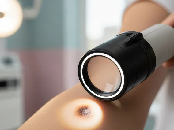
Recognizing Stage I Melanoma: Signs and Symptoms
Recognizing stage 1 melanoma symptoms and signs primarily involves vigilance for changes in existing moles or the appearance of new, unusual skin growths. The most widely recognized guide for identifying potential melanoma is the “ABCDE” rule, which helps individuals and clinicians assess suspicious lesions. Early detection is paramount because melanoma is most curable when caught at this localized stage.
The ABCDEs of melanoma are:
- A – Asymmetry: One half of the mole does not match the other half.
- B – Border Irregularity: The edges are ragged, notched, or blurred.
- C – Color Variation: The mole has uneven color, with shades of brown, black, tan, red, white, or blue.
- D – Diameter: The spot is larger than 6 millimeters (about the size of a pencil eraser), though melanomas can be smaller.
- E – Evolving: The mole is changing in size, shape, color, or elevation, or any new symptoms like bleeding, itching, or crusting appear.
Regular self-skin exams and annual professional skin checks by a dermatologist are crucial for identifying these signs early. Any suspicious lesion should be promptly evaluated by a healthcare professional.
Stage I Melanoma Treatment and Prognosis
The primary stage 1 melanoma treatment options revolve around surgical removal of the tumor. This procedure, known as wide local excision, involves removing the melanoma along with a small margin of surrounding healthy skin to ensure all cancer cells are eliminated. The size of the margin depends on the thickness of the melanoma, typically ranging from 0.5 to 1.0 centimeter for Stage I lesions. For very thin melanomas (T1a), sentinel lymph node biopsy (SLNB) is generally not recommended as the risk of spread to lymph nodes is extremely low. However, for thicker Stage IB melanomas, SLNB may be considered in some cases to check for microscopic spread, although it is often not necessary for Stage I.
The stage 1 melanoma prognosis and survival rate are exceptionally favorable, making it one of the most curable forms of cancer when detected early. According to data from the American Cancer Society, the estimated 5-year survival rate for localized melanoma (which includes Stage I) is approximately 99%. This high survival rate underscores the importance of early diagnosis and prompt treatment. Following surgery, patients typically undergo regular follow-up examinations to monitor for any signs of recurrence or the development of new melanomas. Adherence to sun protection measures, including wearing protective clothing, seeking shade, and using broad-spectrum sunscreen, is also vital for long-term skin health and reducing the risk of future skin cancers.


