Sebaceous Epithelioma
Sebaceous epithelioma is a rare, benign skin tumor originating from sebaceous glands. While generally non-cancerous, understanding its characteristics is crucial for accurate diagnosis and appropriate management.
Key Takeaways
- Sebaceous epithelioma is a rare, benign tumor of the sebaceous glands, typically appearing on the head and neck.
- It often presents as a solitary, firm, yellowish-to-reddish papule or nodule.
- Diagnosis relies heavily on clinical examination and histopathological analysis of a biopsy.
- While benign, surgical excision is the primary and most effective treatment to prevent recurrence and confirm diagnosis.
- It is important to differentiate sebaceous epithelioma from other sebaceous neoplasms, including sebaceous carcinoma, which is malignant.
What is Sebaceous Epithelioma?
Sebaceous epithelioma is a rare, benign adnexal tumor originating from the sebaceous glands of the skin. These glands are responsible for producing sebum, an oily substance that lubricates the skin and hair. Typically, this tumor presents as a solitary, slow-growing lesion, most commonly found on the head and neck, particularly in older adults. While it is generally considered benign, its clinical appearance can sometimes mimic other skin lesions, necessitating careful evaluation. The precise incidence of sebaceous epithelioma is not well-documented due to its rarity, but it is recognized within the spectrum of sebaceous neoplasms.
Identifying Sebaceous Epithelioma: Symptoms, Causes, and Diagnosis
Identifying sebaceous epithelioma symptoms causes and diagnosis involves a combination of clinical observation and histological examination. Clinically, sebaceous epithelioma often appears as a firm, yellowish-to-reddish papule or nodule, ranging from a few millimeters to several centimeters in diameter. The surface may be smooth or slightly lobulated. Patients typically report a painless, slowly enlarging growth. While the exact causes of sebaceous epithelioma are not fully understood, it is believed to arise spontaneously. There is a known association between multiple sebaceous neoplasms, including sebaceous epithelioma, and Muir-Torre syndrome, a rare autosomal dominant disorder characterized by sebaceous tumors and internal malignancies, particularly colorectal cancer. Therefore, if multiple sebaceous lesions are present, screening for Muir-Torre syndrome may be recommended.
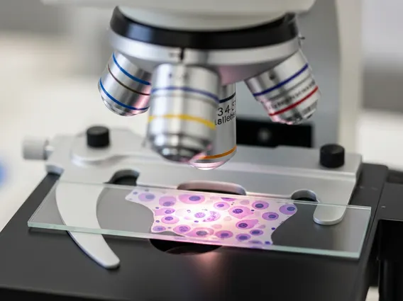
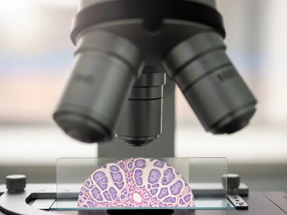
Diagnosing sebaceous epithelioma primarily relies on a skin biopsy followed by histopathological examination. During the biopsy, a small tissue sample of the lesion is removed and analyzed under a microscope by a dermatopathologist. Key microscopic features include nests and cords of basaloid cells with sebaceous differentiation, often surrounded by a prominent stroma. The presence of mature sebocytes and immature sebaceous cells helps distinguish it from other sebaceous tumors. Imaging studies are generally not required for diagnosis unless there is suspicion of deeper invasion or metastasis, which is exceedingly rare for this benign tumor. Differential diagnoses include sebaceous adenoma, basal cell carcinoma with sebaceous differentiation, and sebaceous carcinoma, making accurate histopathological assessment crucial.
Sebaceous Epithelioma Treatment Options
The primary and most effective of the sebaceous epithelioma treatment options is surgical excision. This involves completely removing the tumor with clear margins to prevent recurrence and confirm the benign nature of the lesion through comprehensive histopathological analysis. Given its benign nature, extensive or aggressive surgical procedures are typically not necessary. For smaller lesions, a simple excision is often sufficient. Mohs micrographic surgery may be considered for lesions in cosmetically sensitive areas or those with ill-defined borders, as it allows for precise removal while preserving healthy tissue. Recurrence after complete surgical excision is uncommon.
Other treatment modalities are generally not indicated for sebaceous epithelioma due to its benign characteristic and the effectiveness of surgical removal. These might include cryotherapy or laser ablation, but they are less preferred as they do not allow for complete histopathological evaluation of the excised tissue, which is vital for confirming the diagnosis and ruling out malignancy. Regular follow-up after treatment is usually recommended to monitor for any new lesions, especially in patients with a potential association with Muir-Torre syndrome, where ongoing surveillance for internal malignancies is crucial. Patients are also advised to perform self-skin examinations and seek medical attention for any new or changing skin lesions.