Positron Emission Tomography Magnetic Resonance Imaging Scan
Positron Emission Tomography Magnetic Resonance Imaging (PET/MRI) Scan is a cutting-edge diagnostic tool that combines two powerful imaging technologies into a single examination. This integrated approach provides highly detailed anatomical and metabolic information, offering comprehensive insights into various disease processes.
Key Takeaways
- Positron Emission Tomography Magnetic Resonance Imaging (PET/MRI) Scan is a hybrid imaging modality combining the functional insights of PET with the detailed anatomical imaging of MRI.
- It works by simultaneously acquiring data from a radioactive tracer (for metabolic activity) and strong magnetic fields (for soft tissue detail).
- Primary clinical applications include oncology, neurology, and cardiology, offering precise disease localization and characterization.
- PET MRI scan uses and benefits include superior soft tissue contrast, reduced radiation exposure compared to PET/CT, and comprehensive disease assessment.
- PET MRI vs PET CT scan highlights differences in radiation dose, soft tissue imaging capabilities, and specific clinical advantages.
What is Positron Emission Tomography Magnetic Resonance Imaging (PET/MRI) Scan?
Positron Emission Tomography Magnetic Resonance Imaging (PET/MRI) Scan is an advanced medical imaging technique that integrates the functional imaging capabilities of Positron Emission Tomography (PET) with the high-resolution anatomical imaging of Magnetic Resonance Imaging (MRI). This innovative hybrid system allows clinicians to simultaneously visualize metabolic activity and detailed anatomical structures within the body. By combining these two modalities, PET/MRI provides a more complete picture of disease, enabling earlier detection, more accurate staging, and better monitoring of treatment response, particularly in complex conditions affecting soft tissues.
How PET/MRI Scans Work and Their Clinical Applications
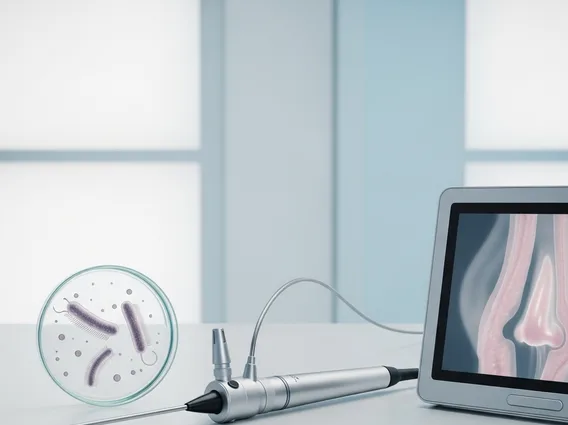
The mechanism of a PET/MRI scan involves the concurrent acquisition of data from both PET and MRI components. The PET component begins with the intravenous injection of a small amount of a radioactive tracer, such as fluorodeoxyglucose (FDG), which is a glucose analog. This tracer accumulates in cells with high metabolic activity, often indicative of cancerous tumors, inflammation, or specific neurological processes. The PET scanner then detects the gamma rays emitted by the tracer, creating images that highlight areas of increased metabolic function.
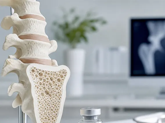
Simultaneously, the MRI component uses strong magnetic fields and radio waves to generate highly detailed images of organs, soft tissues, bone marrow, and blood vessels. Unlike CT scans, MRI does not use ionizing radiation for its anatomical imaging. The synchronized data acquisition of PET and MRI allows for precise co-registration of functional and anatomical images, meaning metabolic abnormalities detected by PET can be accurately localized to specific anatomical structures identified by MRI. This synergy is crucial for accurate diagnosis and staging.
The PET MRI scan uses and benefits are extensive across several medical specialties. Key applications include:
- Oncology: Staging various cancers, assessing tumor response to therapy, detecting recurrence, and guiding biopsies, particularly effective for brain, head and neck, liver, and pelvic cancers.
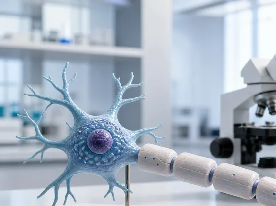
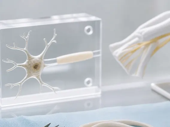
- Neurology: Evaluating brain tumors, epilepsy, dementia, and other neurodegenerative disorders by mapping metabolic changes in the brain.
- Cardiology: Assessing myocardial viability and inflammation in conditions like sarcoidosis or vasculitis.
- Pediatrics: Offering a significant advantage due to reduced radiation exposure, making it safer for children requiring repeated scans.
The benefits include superior soft tissue contrast from MRI, precise anatomical localization of metabolic abnormalities, and a lower overall radiation dose compared to PET/CT, as the MRI component does not use ionizing radiation.
PET/MRI vs. PET/CT: Key Differences and Advantages
When considering PET MRI vs PET CT scan, it is important to understand that both are hybrid imaging technologies designed to combine functional and anatomical information. PET/CT has been the established standard for many years, integrating PET with Computed Tomography (CT). While both modalities are highly effective, they offer distinct advantages and disadvantages depending on the clinical scenario.
The primary difference lies in the anatomical imaging component. PET/CT uses CT, which provides rapid imaging and excellent bone detail, but exposes the patient to additional ionizing radiation. PET/MRI, conversely, uses MRI, which offers superior soft tissue contrast without additional ionizing radiation. This makes PET/MRI particularly advantageous for imaging areas rich in soft tissue, such as the brain, liver, and pelvis, where MRI’s detailed anatomical resolution can better delineate lesions and their relationship to surrounding structures. For instance, in pediatric oncology, where minimizing radiation exposure is critical, PET/MRI is often preferred.
However, PET/CT still holds advantages in certain situations, such as lung imaging where motion artifacts can be more problematic for MRI, or for rapid whole-body scans. The choice between PET/MRI and PET/CT often depends on the specific clinical question, patient characteristics, and availability of the technology. According to the World Health Organization (WHO), medical imaging plays a crucial role in global health, and advancements like PET/MRI contribute to more precise diagnostics, especially in complex diseases like cancer.
Here is a comparison of key features:
| Feature | PET/MRI Scan | PET/CT Scan |
|---|---|---|
| Anatomical Component | Magnetic Resonance Imaging (MRI) | Computed Tomography (CT) |
| Ionizing Radiation (Anatomical) | None | Present |
| Soft Tissue Contrast | Superior | Good |
| Bone Imaging | Good (indirect) | Excellent |
| Image Acquisition Speed | Slower than CT | Faster than MRI |
| Primary Applications | Brain, pelvis, liver, pediatrics, soft tissue tumors | Lung, bone, general oncology, rapid whole-body scans |
| Overall Radiation Dose | Lower (PET tracer only) | Higher (PET tracer + CT) |