Photocoagulation
Photocoagulation is a widely utilized medical procedure, primarily in ophthalmology, that employs focused laser light to treat various retinal conditions. This technique is crucial for preventing vision loss by addressing issues such as abnormal blood vessel growth and leakage.
Key Takeaways
- Photocoagulation is a laser-based medical procedure used to treat eye conditions like diabetic retinopathy and macular degeneration.
- It works by using a precise laser beam to create controlled burns on the retina, sealing leaky blood vessels or destroying abnormal tissue.
- The procedure is typically performed in an outpatient setting, often with local anesthesia and pupil dilation.
- Recovery involves temporary blurred vision and light sensitivity, with specific post-procedure care instructions to ensure optimal healing.
- This treatment aims to preserve existing vision and prevent further damage, significantly impacting patient quality of life.
What is Photocoagulation?
Photocoagulation refers to a therapeutic medical procedure that uses a laser to create precise, controlled burns on tissue. In ophthalmology, this technique is a cornerstone for managing various retinal diseases. The primary goal of photocoagulation is to prevent further vision loss by targeting and treating specific areas of the retina. This can involve sealing leaky blood vessels, destroying abnormal tissue, or creating scar tissue to prevent retinal detachment.
The application of this technique, often referred to as photocoagulation eye treatment explained, is particularly vital for conditions like diabetic retinopathy, a complication of diabetes that damages blood vessels in the light-sensitive tissue at the back of the eye. According to the World Health Organization (WHO), diabetic retinopathy affects approximately one-third of people living with diabetes globally, making treatments like photocoagulation essential for preserving sight. It is also used for certain types of macular degeneration, retinal tears, and other vascular disorders of the eye.
How Photocoagulation Works
The mechanism behind photocoagulation involves the precise delivery of high-energy laser light to the target tissue. When the laser beam strikes the retina, the light energy is absorbed by pigment cells, primarily melanin, and converted into heat. This localized heat causes the tissue to coagulate, essentially creating a tiny, controlled burn or scar. This process has several therapeutic effects:
- Sealing Leaky Vessels: In conditions like diabetic retinopathy, abnormal blood vessels can leak fluid and blood into the retina, causing swelling and vision impairment. The laser seals these vessels, preventing further leakage.
- Destroying Abnormal Tissue: Proliferative diabetic retinopathy involves the growth of new, fragile blood vessels (neovascularization) that can bleed and lead to scar tissue formation. The laser destroys these abnormal vessels.
- Reducing Oxygen Demand: In extensive cases, such as panretinal photocoagulation, numerous small laser spots are applied to the peripheral retina. This reduces the oxygen demand of the treated areas, redirecting oxygen to the more critical central retina (macula) and inhibiting the growth of new, problematic blood vessels.
The laser used is highly focused, allowing ophthalmologists to target specific areas of the retina while minimizing damage to surrounding healthy tissue. Different wavelengths of laser light may be used depending on the specific condition and the depth of penetration required.
Photocoagulation Procedure and Recovery
The photocoagulation procedure and recovery process typically begins with preparation in an outpatient clinic. Patients usually receive dilating eye drops to widen the pupil, allowing the ophthalmologist a clear view of the retina. Anesthetic eye drops are also administered to numb the eye, ensuring comfort during the procedure. In some cases, a contact lens may be placed on the eye to help focus the laser and stabilize the eye.
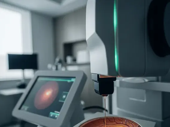
During the procedure, the patient sits in a chair with their chin resting on a support, similar to a standard eye exam. The ophthalmologist then uses a specialized microscope and laser delivery system to apply precise laser pulses to the retina. Patients may see flashes of light and feel a mild stinging or pressure sensation, but significant pain is uncommon due to the anesthetic. The duration of the procedure varies depending on the extent of the condition, ranging from a few minutes to half an hour or more.
After the procedure, patients can typically go home the same day. Recovery involves:
- Blurred Vision: Vision may be temporarily blurred or dim for several hours due to pupil dilation and the laser effects.
- Light Sensitivity: Eyes may be sensitive to light, so wearing sunglasses is often recommended.
- Mild Discomfort: Some patients experience mild eye discomfort, which can usually be managed with over-the-counter pain relievers.
- Activity Restrictions: Strenuous activities might be limited for a short period, as advised by the doctor.
- Follow-up Appointments: Regular follow-up visits are crucial to monitor healing and assess the effectiveness of the treatment.
While photocoagulation aims to preserve existing vision and prevent further deterioration, it generally does not restore vision already lost. The success of the treatment depends on the underlying condition, its severity, and the patient’s adherence to post-procedure care.