
Pet Mri Scan
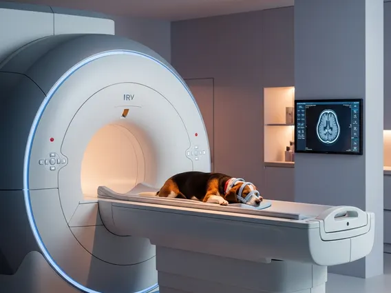
A Pet MRI Scan is an advanced diagnostic imaging technique used in veterinary medicine to create detailed images of an animal’s internal structures. This non-invasive procedure is crucial for diagnosing a wide range of conditions affecting pets.
Key Takeaways
- A Pet MRI Scan is a sophisticated imaging tool for animals, providing highly detailed views of soft tissues, the brain, and the spinal cord.
- It operates using strong magnetic fields and radio waves, making it safe as it does not involve ionizing radiation.
- The primary benefit of pet MRI for diagnosis is its ability to detect subtle abnormalities that other imaging methods might miss, leading to more accurate diagnoses.
- Pets require general anesthesia for the procedure to ensure they remain still and safe throughout the scan.
- Pet MRI scan cost explained varies significantly based on factors like location, the complexity of the scan, and the need for anesthesia.
What is a Pet MRI Scan?
A Pet MRI Scan, or Magnetic Resonance Imaging for pets, is a state-of-the-art diagnostic tool that veterinarians utilize to obtain high-resolution images of an animal’s internal anatomy. Similar to human MRI, it employs a powerful magnetic field and radio waves to generate detailed cross-sectional pictures of organs, soft tissues, bone, and virtually all other internal body structures. This technology is particularly adept at visualizing soft tissues, such as the brain, spinal cord, muscles, and ligaments, making it invaluable for diagnosing neurological, orthopedic, and oncological conditions in companion animals.
The procedure is non-invasive and does not involve ionizing radiation, which is a key advantage over X-rays or CT scans, especially for sensitive tissues. Its ability to differentiate between various soft tissues with exceptional clarity allows veterinarians to identify abnormalities like tumors, inflammation, infections, and degenerative diseases with greater precision. This level of detail is often critical for formulating an accurate diagnosis and developing an effective treatment plan for pets.
How Pet MRI Scans Work for Diagnosis
How do pet MRI scans work involves a complex yet safe process. When a pet undergoes an MRI, it is placed within a strong magnetic field. This field causes the protons within the water molecules in the pet’s body to align temporarily. Short bursts of radio waves are then emitted, which temporarily knock these aligned protons out of alignment. When the radio waves are turned off, the protons relax back into alignment, releasing energy signals in the process. These signals are detected by the MRI scanner and sent to a computer, which translates them into detailed images.
The diagnostic power of this technique lies in its ability to distinguish between different types of tissues based on how quickly their protons realign. For example, diseased tissues often have different water content or cellular structures compared to healthy tissues, leading to variations in signal intensity that are visible on the MRI images. This allows for the detection of:
- Brain and spinal cord disorders (e.g., tumors, herniated discs, inflammation)
- Joint and soft tissue injuries (e.g., ligament tears, muscle damage)
- Certain types of cancer and their extent
- Internal organ abnormalities (though less common than for neurological or orthopedic issues)
The Benefits of pet MRI for diagnosis are significant, offering unparalleled detail for soft tissue structures. Because pets must remain perfectly still for the duration of the scan, which can last from 30 minutes to over an hour, general anesthesia is always required. This ensures both the safety of the animal and the quality of the images obtained. According to a study published in the Journal of the American Veterinary Medical Association, advanced imaging techniques like MRI have significantly improved diagnostic accuracy in veterinary neurology, leading to better outcomes for pets with complex conditions.
Understanding Pet MRI Scan Costs
Pet MRI scan cost explained is a common concern for pet owners, as the procedure represents a significant investment in their pet’s health. The cost can vary widely, typically ranging from several hundred to several thousand dollars, influenced by a number of factors. These include the geographic location of the veterinary hospital or imaging center, the specific area of the body being scanned, and the complexity of the case.
| Factor | Impact on Cost |
|---|---|
| Location | Costs can be higher in urban areas or regions with a higher cost of living. |
| Scan Area | Scanning larger or multiple areas (e.g., entire spine vs. single joint) increases time and cost. |
| Anesthesia | Includes pre-anesthetic blood work, the anesthetic itself, monitoring during the procedure, and recovery. This is a major component of the overall cost. |
| Specialist Interpretation | The images are often interpreted by a board-certified veterinary radiologist, whose expertise adds to the overall fee. |
| Emergency vs. Scheduled | Emergency scans may incur higher fees due to immediate staffing and equipment availability. |
While the cost can be substantial, many pet owners find the diagnostic clarity provided by a Pet MRI Scan to be invaluable. It can prevent unnecessary treatments, guide precise surgical interventions, and ultimately improve the quality of life for their beloved companions. Pet insurance policies often cover a portion of advanced diagnostic procedures like MRI, so it is advisable for pet owners to check their policy details or consider obtaining insurance to help manage potential expenses.


