Perfusion Magnetic Resonance Imaging
Perfusion Magnetic Resonance Imaging (Perfusion MRI) is a specialized diagnostic technique that provides crucial insights into blood flow within tissues and organs. This advanced imaging method plays a vital role in understanding various medical conditions by visualizing the delivery of blood to specific areas of the body.
Key Takeaways
- Perfusion MRI measures blood flow and volume in tissues, offering functional information beyond anatomical details.
- It typically involves the injection of a contrast agent to track its passage through the microvasculature.
- The technique is invaluable for diagnosing and managing conditions like stroke, brain tumors, and cardiac ischemia.
- It helps clinicians assess tissue viability, tumor aggressiveness, and treatment response.
- Understanding Perfusion Magnetic Resonance Imaging is essential for advanced neurological and oncological diagnostics.
What is Perfusion Magnetic Resonance Imaging (MRI)?
Perfusion Magnetic Resonance Imaging (Perfusion MRI) is an advanced medical imaging technique that assesses the amount of blood delivered to a specific region of tissue over time. Unlike conventional MRI, which primarily provides structural information, Perfusion MRI offers functional data by quantifying blood flow, blood volume, and capillary permeability. This capability is critical for evaluating tissue viability and metabolic activity, which can be altered in various disease states. By providing a dynamic view of blood supply, Perfusion MRI helps clinicians gain a deeper understanding of tissue health and disease progression, making it an indispensable tool for diagnostics and treatment planning.
The core principle behind Perfusion MRI involves tracking the passage of a contrast agent through the body’s microvasculature. This allows for the creation of detailed maps that illustrate how well blood is perfusing different tissues. This method is particularly useful for understanding conditions where blood supply is compromised or altered, such as in stroke or cancer. Effective understanding Perfusion Magnetic Resonance Imaging enables medical professionals to make more informed decisions regarding patient care, from initial diagnosis to monitoring therapeutic efficacy.
How Does Perfusion MRI Work?
Perfusion MRI operates by detecting changes in the magnetic properties of tissue as a contrast agent, typically a gadolinium-based compound, passes through the bloodstream. There are primarily two main techniques used: Dynamic Susceptibility Contrast (DSC) MRI and Dynamic Contrast-Enhanced (DCE) MRI.
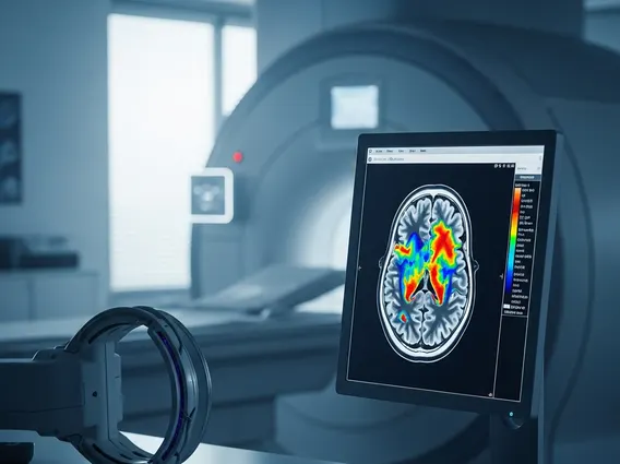
In DSC-MRI, a bolus of contrast agent is rapidly injected intravenously. As the contrast agent passes through the capillaries, it causes a temporary decrease in the T2* signal intensity of the surrounding tissue. MRI scanners capture a series of rapid images during this first pass, and specialized software then analyzes these signal changes over time. From this data, quantitative maps of cerebral blood flow (CBF), cerebral blood volume (CBV), and mean transit time (MTT) can be generated. These parameters reflect the efficiency and volume of blood delivery to the tissue.
DCE-MRI, on the other hand, measures the leakage of contrast agent from the vasculature into the extravascular-extracellular space. This technique is more sensitive to capillary permeability and can provide insights into the integrity of the blood-brain barrier or tumor angiogenesis. By tracking the enhancement patterns over a longer period, DCE-MRI can quantify parameters such as Ktrans (transfer constant) and Ve (extravascular-extracellular volume fraction), which are crucial for assessing tumor characteristics and response to anti-angiogenic therapies.
Clinical Applications of Perfusion MRI
Perfusion MRI has a wide range of clinical applications, significantly enhancing diagnostic capabilities across several medical specialties. Its ability to visualize and quantify blood flow makes it invaluable for assessing conditions where tissue perfusion is critical. The insights gained from perfusion MRI applications help guide treatment decisions and monitor patient outcomes.
Key clinical applications include:
- Stroke Management: Perfusion MRI is crucial in acute stroke for identifying the ischemic penumbra—tissue that is at risk but potentially salvageable. This helps determine which patients will benefit most from reperfusion therapies, such as thrombolysis or thrombectomy. According to the World Health Organization (WHO), stroke is a leading cause of death and disability globally, and timely diagnosis with tools like Perfusion MRI is vital for improving patient prognosis.
- Brain Tumor Characterization: It helps differentiate between high-grade and low-grade brain tumors, distinguish tumor recurrence from radiation necrosis, and assess the effectiveness of chemotherapy or radiation therapy by monitoring changes in tumor vascularity.
- Neurodegenerative Diseases: While less common, it can sometimes be used to study altered perfusion patterns in conditions like Alzheimer’s disease, providing insights into regional brain activity.
- Cardiac Imaging: In cardiology, Perfusion MRI can detect myocardial ischemia by identifying areas of reduced blood flow to the heart muscle, often in response to stress. This helps diagnose coronary artery disease and assess its severity.
These diverse applications underscore the utility of Perfusion MRI as a powerful diagnostic tool, offering functional information that complements traditional anatomical imaging and significantly impacts patient management.