Lymphangiography
Lymphangiography is a specialized medical imaging procedure designed to visualize the lymphatic system. It involves injecting a contrast agent into lymphatic vessels, allowing healthcare professionals to assess their structure and function, which is crucial for diagnosing various conditions affecting this vital part of the immune system.
Key Takeaways
- Lymphangiography is a diagnostic imaging technique that uses a contrast dye to visualize lymphatic vessels and nodes.
- The procedure typically involves injecting a specialized dye into lymphatic vessels, often in the foot, followed by X-ray imaging.
- It is primarily used to diagnose lymphatic system disorders, identify cancer spread, and guide therapeutic interventions.
- Patients should expect preparation steps, the injection process, imaging, and post-procedure monitoring.
- Potential risks include skin discoloration, allergic reactions, and localized discomfort, which are generally manageable.
What is Lymphangiography?
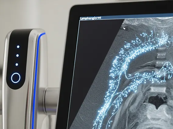
Lymphangiography refers to a medical imaging technique used to obtain detailed images of the lymphatic system, including lymphatic vessels and lymph nodes. This diagnostic procedure involves the injection of a special contrast dye, typically oil-based, directly into a lymphatic vessel. Once injected, the dye travels through the lymphatic system, making the otherwise transparent vessels and nodes visible on X-ray images, computed tomography (CT) scans, or magnetic resonance imaging (MRI).
The primary purpose of Lymphangiography is to identify abnormalities within the lymphatic system. This can include blockages, leaks, malformations, or the presence of cancerous cells that have spread to the lymph nodes. By providing a clear map of the lymphatic pathways, it aids clinicians in making accurate diagnoses and planning appropriate treatment strategies for conditions affecting lymphatic flow and immune function.
The Lymphangiography Procedure: What to Expect
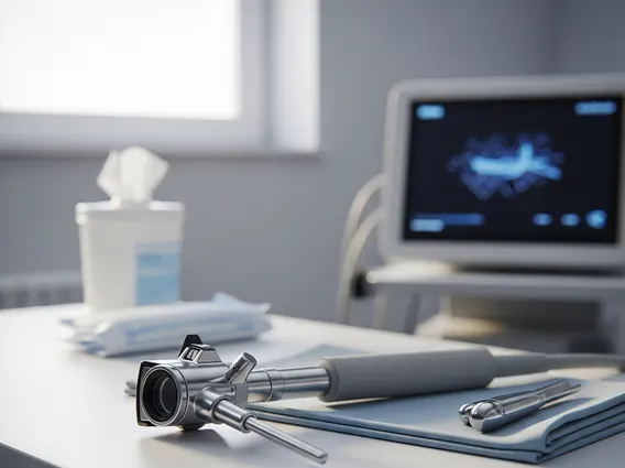
The lymphangiography procedure explained involves several steps, typically performed in a hospital or specialized imaging center. Patients are usually advised to fast for a few hours before the procedure and to inform their healthcare provider of any allergies, especially to iodine or contrast dyes. The process generally begins with the patient lying comfortably on an examination table.
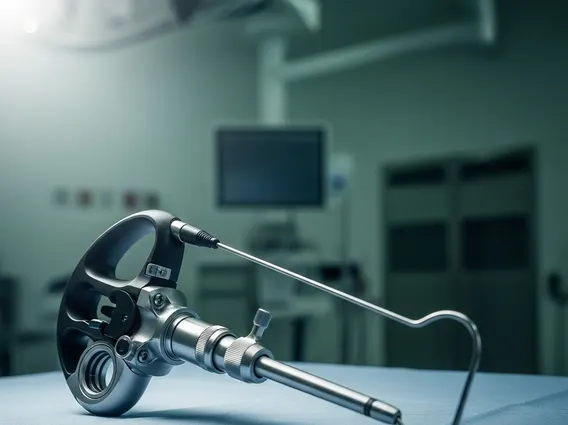
A small amount of local anesthetic is applied to numb the skin, often on the top of the foot. A very small incision is then made to locate a lymphatic vessel, into which a thin catheter is inserted. The contrast dye is slowly injected through this catheter, which can take several hours as the dye gradually fills the lymphatic system. During and after the injection, a series of X-rays or other imaging scans are taken to track the dye’s movement and visualize the lymphatic structures. The dye remains in the lymphatic system for an extended period, sometimes days or weeks, allowing for delayed imaging to assess lymphatic function over time. After the procedure, the catheter is removed, and the small incision is closed.
Patients may experience some discomfort or a feeling of pressure during the injection. Post-procedure, it is common for urine, stool, or skin to have a bluish tint for a few days due to the dye. Patients are typically monitored for a short period before being discharged with instructions for wound care and activity restrictions.
Uses and Potential Side Effects of Lymphangiography
The uses of lymphangiography are diverse, primarily focusing on the diagnosis and management of lymphatic system disorders. It is particularly valuable in cases where other imaging techniques may not provide sufficient detail. Key applications include:
- Diagnosing Lymphedema: Identifying blockages or damage in lymphatic vessels that cause swelling.
- Detecting Lymphatic Leaks: Pinpointing the source of chylous effusions (fluid leaks containing lymph) in the chest or abdomen.
- Assessing Cancer Spread: Determining if certain cancers, such as lymphoma or melanoma, have spread to the lymph nodes.
- Guiding Interventions: Assisting in the planning of surgical procedures or targeted therapies for lymphatic conditions.
- Evaluating Lymphatic Malformations: Visualizing congenital abnormalities of the lymphatic vessels.
While generally safe, there are potential lymphangiography side effects that patients should be aware of. Common side effects include temporary skin discoloration (often a bluish tint) at the injection site or elsewhere on the body, which typically fades over time. Some individuals may experience localized pain, tenderness, or swelling at the injection site. More serious, though less common, side effects can include allergic reactions to the contrast dye, ranging from mild rashes to severe anaphylaxis. Other potential complications include infection at the injection site, extravasation (leakage of dye into surrounding tissues), or, rarely, damage to the lymphatic vessels. Healthcare providers carefully weigh these risks against the diagnostic benefits for each patient.