Intravenous Urography
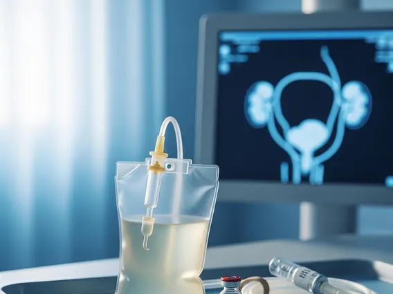
Intravenous Urography (IVU), also known as an intravenous pyelogram (IVP), is a diagnostic imaging test that uses X-rays and a special contrast dye to visualize the kidneys, ureters, and bladder. This procedure helps medical professionals assess the structure and function of the urinary tract.
Key Takeaways
- Intravenous Urography (IVU) is an X-ray imaging test using contrast dye to examine the urinary system.
- It helps diagnose conditions like kidney stones, tumors, and blockages in the kidneys, ureters, and bladder.
- Preparation involves fasting and discussing medications with your doctor.
- The procedure includes intravenous contrast injection followed by a series of X-rays.
- Potential risks include allergic reactions to the dye and radiation exposure, which are generally low but require consideration.
What is Intravenous Urography?
Intravenous Urography (IVU), also known as an intravenous pyelogram (IVP), is a specialized radiological examination designed to evaluate the entire urinary tract. This diagnostic procedure involves injecting a contrast material, typically iodine-based, into a vein. As the contrast agent travels through the bloodstream, it is filtered by the kidneys and excreted into the urine, making the kidneys, ureters (tubes connecting kidneys to the bladder), and bladder visible on X-ray images. The primary purpose of IVU is to assess the anatomical structure and functional integrity of these organs, helping to identify abnormalities that might not be visible with standard X-rays.
IVU is particularly useful for detecting a range of conditions affecting the urinary system. It provides detailed images of the internal structures, allowing clinicians to observe how urine flows through the system. This comprehensive view aids in diagnosing issues such as kidney stones, tumors, cysts, and blockages. While other imaging modalities like CT scans and MRI are now more common for some conditions, IVU remains a valuable tool in specific clinical scenarios, especially for visualizing the collecting system of the kidneys and the ureters.
Intravenous Urography Procedure and Preparation
The Intravenous urography procedure explained involves several key steps, beginning with thorough patient preparation to ensure accurate and safe imaging. Before the procedure, patients are typically instructed to fast for several hours (usually 8-12 hours) to ensure the bladder is empty and to minimize the risk of nausea from the contrast dye. It is also crucial to discuss all current medications, allergies, and medical history with the healthcare provider, especially regarding kidney function and any previous reactions to contrast agents. Patients may be advised to adjust certain medications, such as metformin, which can interact with iodine-based contrast in individuals with impaired kidney function.
During the procedure, the patient lies on an X-ray table. A healthcare professional will inject the iodine-based contrast dye into a vein, usually in the arm. A series of X-ray images are then taken at specific intervals as the contrast material moves through the kidneys, ureters, and into the bladder. This allows radiologists to track the dye’s progression and identify any obstructions or structural anomalies. Additional images may be taken after the patient urinates to assess bladder emptying. The entire process typically takes about 30 to 60 minutes, though it can sometimes extend longer depending on the findings.
- Fasting for 8-12 hours before the procedure.
- Reviewing all medications, especially those affecting kidney function.
- Informing the medical team about any allergies, particularly to iodine or contrast dyes.
- Ensuring adequate hydration prior to fasting.
Uses, Risks, and Patient Guidance for Intravenous Urography
The Intravenous urography uses and risks are important considerations for both patients and clinicians. IVU is primarily utilized to diagnose and evaluate various conditions affecting the urinary tract. Common uses include identifying the location and size of kidney stones, detecting tumors or cysts in the kidneys or bladder, and assessing for blockages in the ureters that might be causing hydronephrosis (swelling of a kidney due to urine backup). It can also help diagnose congenital anomalies of the urinary system, such as a duplicated collecting system, and evaluate injuries to the kidneys or bladder following trauma.
While generally safe, IVU carries certain risks. The most common risk is an allergic reaction to the iodine-based contrast dye, which can range from mild symptoms like hives or itching to more severe reactions such as difficulty breathing or anaphylaxis. Patients with a history of allergies, asthma, or previous reactions to contrast are at a higher risk. Another concern is the potential for contrast-induced nephropathy, a temporary or, rarely, permanent decline in kidney function, particularly in individuals with pre-existing kidney disease. Radiation exposure is also a factor, though the dose is typically low and considered safe for most diagnostic purposes. According to the American College of Radiology, the benefits of diagnostic imaging generally outweigh the risks of radiation exposure when medically indicated.
For those undergoing the procedure, comprehensive Intravenous urography patient guide ensures a smooth experience. After the IVU, patients are usually encouraged to drink plenty of fluids to help flush the contrast dye out of their system. It is important to monitor for any delayed allergic reactions, such as skin rashes or itching, and to report them to a healthcare provider. Patients should contact their doctor immediately if they experience severe pain, fever, difficulty urinating, or any signs of a serious allergic reaction after returning home. Following post-procedure instructions carefully is crucial for recovery and to mitigate potential complications.