Intravenous Pyelogram
An Intravenous Pyelogram (IVP) is a specialized X-ray examination used to visualize the kidneys, ureters, and bladder. This diagnostic procedure helps identify abnormalities within the urinary tract.
Key Takeaways
- Intravenous Pyelogram (IVP) is an imaging test that uses a contrast dye injected into a vein to highlight the urinary system on X-rays.
- The procedure involves preparation, dye injection, and a series of X-rays to track the dye’s path through the kidneys, ureters, and bladder.
- IVP is primarily used to diagnose conditions such as kidney stones, tumors, blockages, and congenital abnormalities of the urinary tract.
- Potential risks include allergic reactions to the contrast dye, kidney damage, and radiation exposure, which are carefully managed by healthcare professionals.
- Interpreting IVP results involves assessing the flow of the contrast, the structure of the organs, and identifying any obstructions or unusual growths.
What is Intravenous Pyelogram (IVP)?
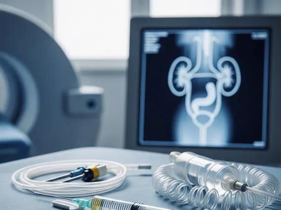
An Intravenous Pyelogram (IVP) is a radiological imaging technique that uses X-rays and a special contrast material to examine the kidneys, ureters, and bladder. This diagnostic tool is crucial for evaluating the structure and function of the urinary system. During the procedure, a water-soluble iodine-based contrast dye is injected into a vein, typically in the arm. As the kidneys filter this dye from the bloodstream, it travels through the urinary tract, making these structures visible on X-ray images. This allows healthcare providers to detect and assess various conditions affecting the urinary system that might not be visible on standard X-rays.
The primary purpose of an IVP is to help diagnose issues such as kidney stones, blockages in the urinary tract, tumors, cysts, and congenital abnormalities. It provides detailed images that can reveal the size, shape, and position of the kidneys, as well as the patency and integrity of the ureters and bladder. The contrast dye helps outline these organs, making it easier to identify any structural changes or obstructions that could be causing symptoms like pain, blood in the urine, or recurrent urinary tract infections.
Intravenous Pyelogram Procedure: What to Expect
The intravenous pyelogram procedure explained typically begins with preparation instructions from your doctor, which may include fasting for several hours before the exam and possibly taking a laxative to clear the bowels, ensuring clearer images. Upon arrival at the imaging facility, you will be asked to change into a hospital gown and lie on an X-ray table. A healthcare professional will then insert an intravenous (IV) line, usually into a vein in your arm, through which the contrast dye will be injected.
As the contrast material enters your bloodstream, you might experience a warm flush throughout your body, a metallic taste in your mouth, or a temporary sensation of nausea. These sensations are usually brief and normal. A series of X-ray images will be taken at different time intervals as the dye travels through your kidneys, ureters, and bladder. This allows the radiologist to observe how well your kidneys are filtering the dye and how it flows through the urinary system. You may be asked to change positions or hold your breath briefly during some X-rays. The entire procedure usually takes about 30 to 60 minutes, though it can sometimes extend longer if delayed images are needed to track the dye’s progression.
- Before the procedure: Fasting for several hours, possibly bowel preparation.
- During the procedure: IV contrast injection, series of X-rays at intervals.
- Sensations: Warm flush, metallic taste, temporary nausea.
- Duration: Typically 30-60 minutes.
Uses, Risks, and Interpreting IVP Results
The intravenous pyelogram uses and risks are important considerations for patients and clinicians. IVP is widely used to diagnose a range of conditions affecting the urinary system. Common uses include identifying kidney stones, detecting tumors or cysts in the kidneys or bladder, evaluating for blockages in the ureters, and assessing for congenital abnormalities of the urinary tract. It can also help investigate causes of blood in the urine (hematuria) or flank pain. However, like all medical procedures, IVP carries certain risks. The primary risks involve allergic reactions to the iodine-based contrast dye, which can range from mild (hives, itching) to severe (difficulty breathing, anaphylaxis). There is also a risk of kidney damage, particularly in individuals with pre-existing kidney disease, and exposure to ionizing radiation, which is generally low but cumulative.
Understanding the intravenous pyelogram results meaning is crucial for diagnosis and treatment planning. After the images are taken, a radiologist, a doctor specializing in interpreting medical images, will analyze them. They will look for several key indicators, including the size, shape, and position of the kidneys, the presence of any stones or calcifications, and the smooth flow of the contrast dye through the ureters to the bladder. Abnormal findings might include: obstructions (such as kidney stones or tumors) that block the flow of urine, structural anomalies, or masses that indicate growths. The radiologist will then provide a detailed report to your referring physician, who will discuss the findings with you and determine the appropriate next steps, which may include further testing or treatment.