Cytogenetic Analysis
Cytogenetic Analysis is a specialized laboratory technique used to study chromosomes, the structures within cells that contain our genetic material. This analysis plays a crucial role in identifying chromosomal abnormalities that can lead to various medical conditions, from developmental disorders to cancer.

Key Takeaways
- Cytogenetic Analysis examines chromosomes for numerical or structural changes.
- It utilizes techniques like karyotyping, FISH, and chromosomal microarray to visualize genetic material.
- This analysis is vital for diagnosing genetic disorders in prenatal and postnatal settings.
- It significantly aids in cancer diagnosis, prognosis, and monitoring treatment effectiveness.
- Understanding chromosomal changes helps guide patient management and genetic counseling.
What is Cytogenetic Analysis?
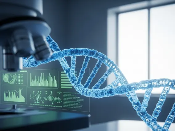
Cytogenetic Analysis refers to the study of chromosomes to detect structural or numerical abnormalities. Chromosomes, which are thread-like structures located inside the nucleus of animal and plant cells, carry genetic information in the form of DNA. By examining these structures, scientists and clinicians can identify changes that may be associated with genetic disorders, developmental delays, infertility, and certain types of cancer. This diagnostic tool provides critical insights into an individual’s genetic makeup, helping to explain the underlying causes of various medical conditions.
The primary goal of cytogenetic analysis is to visualize and evaluate the entire set of chromosomes, known as a karyotype, from a patient’s cells. This allows for the detection of extra or missing chromosomes (numerical abnormalities) or rearrangements within chromosomes (structural abnormalities), such as translocations, inversions, or deletions. Such abnormalities can have significant health implications, influencing growth, development, and disease susceptibility.
How Cytogenetic Analysis Works and Its Techniques
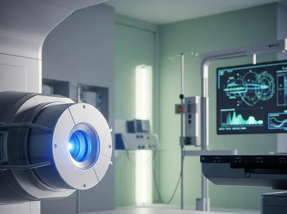
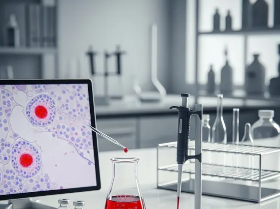
The process of how does cytogenetic analysis work typically begins with obtaining a sample of cells, which can come from various sources such as blood, bone marrow, amniotic fluid, chorionic villi, or solid tumor biopsies. These cells are then cultured in a laboratory to stimulate cell division. During a specific phase of cell division (metaphase), chromosomes become condensed and visible under a microscope. The cells are then harvested, treated to swell and spread the chromosomes, and stained to reveal characteristic banding patterns unique to each chromosome.
Several types of cytogenetic analysis techniques are employed, each offering distinct advantages for detecting different kinds of chromosomal aberrations. These techniques include:
- Karyotyping: This is the traditional method where chromosomes are photographed, arranged into a standard format (karyogram), and visually inspected for numerical and large structural changes. It provides a comprehensive overview of the entire chromosome set.
- Fluorescence In Situ Hybridization (FISH): FISH uses fluorescently labeled DNA probes that bind to specific regions on chromosomes. This technique allows for the detection of smaller deletions, duplications, or translocations that might be missed by standard karyotyping, and it can be applied to both dividing and non-dividing cells.
- Chromosomal Microarray (CMA): CMA is a high-resolution technique that can detect very small gains or losses of chromosomal material (copy number variants) across the entire genome, often at a much higher resolution than karyotyping or even some FISH panels. It is particularly useful for identifying cryptic imbalances associated with developmental disorders.
These techniques provide complementary information, and the choice of method depends on the clinical indication and the type of abnormality suspected. For instance, karyotyping is excellent for detecting aneuploidies, while FISH and CMA offer more detailed insights into submicroscopic changes.
Applications of Cytogenetic Analysis
The cytogenetic analysis uses and applications are extensive across various medical fields, making it an indispensable tool in modern medicine. One of its primary applications is in prenatal diagnosis, where it helps identify chromosomal abnormalities in fetuses, such as Down syndrome (Trisomy 21), Edwards syndrome (Trisomy 18), or Patau syndrome (Trisomy 13). This information is crucial for expectant parents and healthcare providers in making informed decisions regarding pregnancy management. According to the World Health Organization (WHO), congenital anomalies affect an estimated 1 in 33 infants globally each year, highlighting the importance of diagnostic tools like cytogenetic analysis.
In postnatal diagnosis, cytogenetic analysis is used to investigate the genetic basis of developmental delays, intellectual disabilities, congenital anomalies, and unexplained growth problems in children. For adults, it helps in evaluating cases of infertility or recurrent miscarriages, as chromosomal rearrangements in one or both partners can lead to reproductive difficulties. Furthermore, in oncology, cytogenetic analysis is vital for diagnosing specific types of cancer, particularly leukemias and lymphomas, by identifying characteristic chromosomal translocations or deletions. These findings can influence prognosis and guide targeted treatment strategies, allowing for more personalized and effective patient care.