Computed Tomography Scan
A Computed Tomography Scan, commonly known as a CT scan, is a sophisticated medical imaging technique that provides detailed cross-sectional images of the body. This non-invasive procedure plays a crucial role in diagnosing a wide range of medical conditions.
Key Takeaways
- A Computed Tomography (CT) scan utilizes X-rays and computer processing to generate highly detailed, cross-sectional images of internal body structures.
- CT scans are essential diagnostic tools for identifying diseases, guiding medical procedures, and monitoring treatment efficacy across various medical specialties.
- The imaging process involves a scanner that rotates around the patient, capturing multiple X-ray views which are then reconstructed into comprehensive images.
- Key benefits include its ability to provide rapid, high-resolution images of bones, soft tissues, and blood vessels, aiding in precise and timely medical assessments.
- Potential considerations include exposure to ionizing radiation and, in some cases, risks associated with contrast agents, such as allergic reactions or kidney impact.
What is a Computed Tomography (CT) Scan?
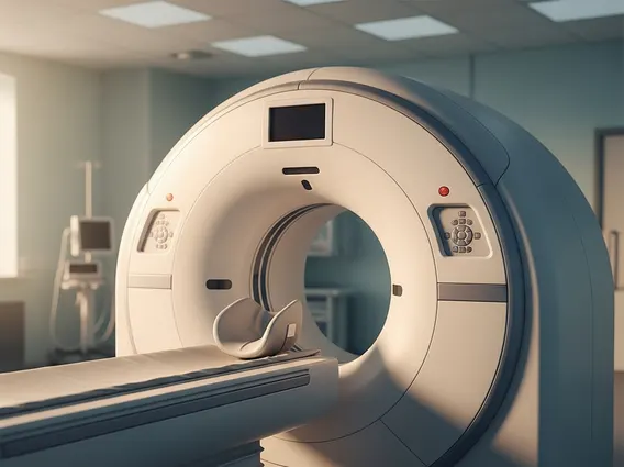
A Computed Tomography Scan (CT scan) is an advanced diagnostic imaging procedure that combines a series of X-ray images taken from different angles around your body and uses computer processing to create cross-sectional (slice) images of the bones, blood vessels, and soft tissues inside your body. Unlike a conventional X-ray, which produces a single, flat image, a CT scan generates detailed views that allow medical professionals to see inside organs and structures from multiple perspectives. This capability makes it an invaluable tool for detecting and characterizing various medical conditions that might not be visible on standard X-rays.
The primary purpose of a Computed Tomography Scan is to provide highly detailed anatomical information, aiding in the accurate diagnosis, staging, and monitoring of diseases. It offers superior clarity for certain tissues and structures, such as complex bone fractures, internal organ injuries, and tumors, compared to other imaging modalities.
How Computed Tomography Scans Work
The mechanism behind a Computed Tomography Scan involves a specialized X-ray unit that rotates around the patient, who lies on a motorized table. As the X-ray tube rotates, it emits a narrow beam of X-rays that passes through the body. On the opposite side of the patient, an array of detectors measures the X-ray beams that have been attenuated (weakened) after passing through different tissues. Denser tissues, like bone, absorb more X-rays, while softer tissues allow more X-rays to pass through.
A powerful computer then processes these thousands of individual X-ray measurements. It reconstructs them into detailed two-dimensional cross-sectional images, or “slices,” of the body. These slices can be viewed individually or stacked together to create three-dimensional representations of organs and structures. In some cases, a contrast material—a special dye—is administered orally or intravenously to enhance the visibility of specific tissues, blood vessels, or organs, allowing for clearer differentiation between healthy and diseased areas.
Uses, Benefits, and Potential Risks of CT Scans
The applications of CT scans are extensive across numerous medical specialties. The uses and benefits of CT scans include their critical role in diagnosing a wide array of conditions, guiding medical procedures, and monitoring treatment responses. For instance, CT scans are frequently used to detect and characterize tumors, assess internal injuries from trauma, diagnose cardiovascular diseases, and identify infections or inflammation. Their ability to provide rapid, high-resolution images makes them indispensable in emergency medicine, where quick and accurate diagnoses are paramount.
Here are some common applications of CT scans:
- Cancer Detection and Staging: Identifying tumors, determining their size and location, and assessing if cancer has spread.
- Trauma Assessment: Quickly evaluating internal injuries, bleeding, and bone fractures after accidents.
- Cardiovascular Imaging: Visualizing blood vessels and assessing conditions like aneurysms or blockages.
- Guiding Procedures: Assisting in biopsies, drainages, and radiation therapy planning by providing real-time anatomical guidance.
- Diagnosing Spinal and Joint Problems: Offering detailed views of bone and soft tissue structures for conditions like herniated discs or arthritis.
While highly beneficial, it is also important to consider the risks and side effects of CT scans. The primary concern is exposure to ionizing radiation. Medical professionals adhere to the “as low as reasonably achievable” (ALARA) principle, ensuring the lowest possible radiation dose is used while maintaining diagnostic image quality. The cumulative effect of multiple CT scans over a lifetime is a consideration, particularly for younger patients. According to the American College of Radiology (ACR), the benefits of a medically indicated CT scan generally outweigh the potential risks.
Another potential risk involves allergic reactions to contrast agents, which can range from mild (hives, itching) to severe (difficulty breathing, anaphylaxis). Patients with kidney impairment may also face a risk of contrast-induced nephropathy, where the contrast material can temporarily worsen kidney function. Therefore, medical history and kidney function are typically assessed before administering contrast. Pregnant women are generally advised to avoid CT scans unless absolutely necessary, due to potential risks to the fetus from radiation exposure.