
Endorectal Ultrasound
Endorectal Ultrasound is a specialized diagnostic imaging technique that provides detailed views of the rectum and surrounding structures. It plays a crucial role in the assessment and staging of various conditions affecting this area, particularly certain types of cancer.
Key Takeaways
- Endorectal Ultrasound (ERUS) uses a small, high-frequency transducer inserted into the rectum to create detailed images.
- The procedure is minimally invasive and typically performed on an outpatient basis.
- ERUS is highly effective for staging rectal cancer, assessing tumor depth, and identifying lymph node involvement.
- It also aids in diagnosing other conditions like perirectal abscesses and inflammatory bowel disease complications.
- The detailed images provided by ERUS are vital for accurate diagnosis and guiding treatment decisions.
What is Endorectal Ultrasound?
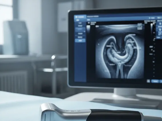
Endorectal Ultrasound (ERUS) is a medical imaging technique that utilizes sound waves to create detailed pictures of the rectum and nearby tissues. A small, specialized ultrasound probe, known as a transducer, is gently inserted into the rectum. This transducer emits high-frequency sound waves that bounce off internal organs and structures, and the echoes are then converted into real-time images displayed on a monitor. This allows healthcare providers to visualize the layers of the rectal wall, assess the depth of any abnormalities, and examine adjacent structures such as lymph nodes and surrounding soft tissues.
The primary advantage of ERUS lies in its ability to provide high-resolution images of the rectal wall, which is often difficult to achieve with external imaging methods. This detailed visualization is critical for distinguishing between different tissue types and identifying subtle changes that may indicate disease. It is a safe, non-invasive procedure that typically causes minimal discomfort and does not involve exposure to radiation.
The Endorectal Ultrasound Procedure
The endorectal ultrasound procedure is generally performed in an outpatient setting and requires minimal preparation. Patients are usually asked to perform a bowel preparation beforehand, similar to what is done for a colonoscopy, to ensure the rectum is clear for optimal imaging. During the procedure, the patient typically lies on their side with knees drawn towards the chest.
A small, lubricated ultrasound probe, which is slightly larger than a finger, is then carefully inserted into the rectum. The probe is slowly advanced and rotated to capture images from different angles. The procedure usually takes about 15 to 30 minutes. Patients may experience a sensation of pressure or fullness, but it is generally not painful. The images are viewed in real-time by the physician, who can assess the anatomy and any abnormalities. After the procedure, patients can typically resume their normal activities immediately.
Uses and Diagnostic Applications of Endorectal Ultrasound
The uses of endorectal ultrasound are diverse, primarily focusing on the detailed assessment of conditions affecting the rectum and surrounding areas. One of its most significant applications is in the staging of rectal cancer. ERUS can accurately determine the depth of tumor invasion into the rectal wall and assess whether the cancer has spread to nearby lymph nodes, which is crucial for guiding treatment decisions such as surgery, radiation, or chemotherapy. According to the American Cancer Society, accurate staging is vital for determining the most effective treatment plan and predicting prognosis for rectal cancer patients.
Beyond cancer staging, ERUS is also invaluable for endorectal ultrasound for diagnosis of other conditions. These include:
- Benign Rectal Tumors: Differentiating between benign and malignant lesions, and assessing their size and location.
- Perirectal Abscesses and Fistulas: Identifying the presence, extent, and tracts of infections or abnormal connections in the perirectal area.
- Inflammatory Bowel Disease (IBD) Complications: Evaluating the extent of inflammation, strictures, or abscesses associated with conditions like Crohn’s disease or ulcerative colitis affecting the rectum.
- Fecal Incontinence: Assessing the integrity of the anal sphincter muscles to identify potential causes of incontinence.
The detailed, cross-sectional images provided by ERUS allow clinicians to make precise diagnoses and plan appropriate interventions, significantly impacting patient management and outcomes.


